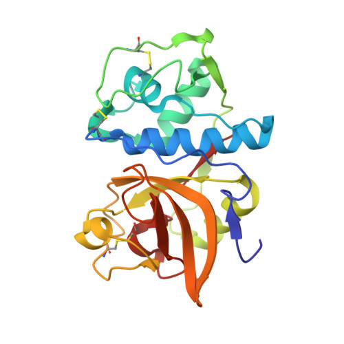
2ATO

Crystal structure of Human Cathepsin K in complex with myocrisin


Experimental Data Snapshot

  • Method: X-RAY DIFFRACTION
  • Resolution: 2.00 Å
  • R-Value Free: 0.198 
  • R-Value Work: 0.197 
  • R-Value Observed: 0.197 

Starting Model: experimental
View more details

wwPDB Validation   3D Report Full Report


Ligand Structure Quality Assessment 


This is version 1.4 of the entry. See complete history


Literature

Effects of disease-modifying anti-rheumatic drugs (DMARDs) on the activities of rheumatoid arthritis-associated cathepsins K and S.

Weidauer, E.Yasuda, Y.Biswal, B.K.Cherny, M.James, M.N.Bromme, D.

(2007) Biol Chem 388: 331-336

  • DOI: https://doi.org/10.1515/BC.2007.037
  • Primary Citation of Related Structures:  
    2ATO

  • PubMed Abstract: 

    Rheumatoid arthritis is an inflammatory and disabling joint disease affecting 0.5-1.5% of the population. Although various anti-inflammatory (NSAIDs) and disease-modifying (DMARDs) drugs are in clinical use, their precise mechanisms of action are not always defined. In this report, we discuss the effects of widely used DMARDs such as gold derivatives and chloroquine on cathepsins K and S, which have been implicated as critical mediators of inflammation and joint erosion in rheumatoid arthritis. We demonstrate that clinically potent gold derivatives inhibit cathepsins K and S in in vitro and cell-based assays. An X-ray analysis of the gold thiomalate/cathepsin K complex reveals that the inhibitor is bound to the active-site cysteine residue of the protease. Chloroquine, a lysosomotropic agent of lower clinical potency than gold derivatives, inhibits neutral pH-labile cathepsins intracellularly, but does not affect the neutral pH-stable cathepsin S. The potent inhibition of cathepsins implicated in the pathogenesis of rheumatoid arthritis by gold derivatives may explain the therapeutic efficacy of these drugs.


  • Organizational Affiliation

    Department of Human Genetics, Mount Sinai School of Medicine, New York, NY 10029, USA.


Macromolecules
Find similar proteins by:  (by identity cutoff)  |  3D Structure
Entity ID: 1
MoleculeChains Sequence LengthOrganismDetailsImage
Cathepsin K215Homo sapiensMutation(s): 0 
Gene Names: CTSKCTSOCTSO2
EC: 3.4.22.38
UniProt & NIH Common Fund Data Resources
Find proteins for P43235 (Homo sapiens)
Explore P43235 
Go to UniProtKB:  P43235
PHAROS:  P43235
GTEx:  ENSG00000143387 
Entity Groups  
Sequence Clusters30% Identity50% Identity70% Identity90% Identity95% Identity100% Identity
UniProt GroupP43235
Sequence Annotations
Expand
  • Reference Sequence
Experimental Data & Validation

Experimental Data

  • Method: X-RAY DIFFRACTION
  • Resolution: 2.00 Å
  • R-Value Free: 0.198 
  • R-Value Work: 0.197 
  • R-Value Observed: 0.197 
  • Space Group: P 21 21 21
Unit Cell:
Length ( Å )Angle ( ˚ )
a = 32.063α = 90
b = 67.398β = 90
c = 93.785γ = 90
Software Package:
Software NamePurpose
CNSrefinement
Blu-Icedata collection
SCALEPACKdata scaling
CNSphasing

Structure Validation

View Full Validation Report


Ligand Structure Quality Assessment 


Entry History 

Deposition Data

Revision History  (Full details and data files)

  • Version 1.0: 2006-08-29
    Type: Initial release
  • Version 1.1: 2008-04-30
    Changes: Version format compliance
  • Version 1.2: 2011-07-13
    Changes: Version format compliance
  • Version 1.3: 2023-08-23
    Changes: Data collection, Database references, Derived calculations, Refinement description
  • Version 1.4: 2024-10-30
    Changes: Structure summary