2BHT

Crystal structure of O-acetylserine sulfhydrylase B


Experimental Data Snapshot

  • Method: X-RAY DIFFRACTION
  • Resolution: 2.10 Å
  • R-Value Free: 
    0.244 (Depositor), 0.230 (DCC) 
  • R-Value Work: 
    0.240 (DCC) 
  • R-Value Observed: 
    0.225 (Depositor) 

Starting Model: experimental
View more details

wwPDB Validation   3D Report Full Report


Ligand Structure Quality Assessment 

Created with Raphaël 2.3.0Worse 01 BetterLigand structure goodness of fit to experimental dataBest fitted PLPClick on this verticalbar to view details

This is version 1.2 of the entry. See complete history


Literature

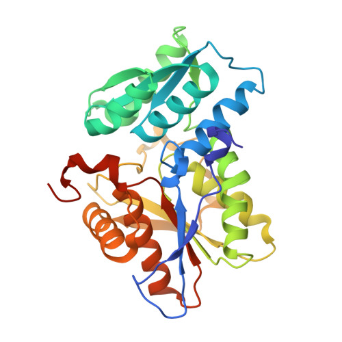
Structure of the O-Acetylserine Sulfhydrylase Isoenzyme Cysm from Escherichia Coli

Claus, M.T.Zocher, G.E.Maier, T.H.P.Schulz, G.E.

(2005) Biochemistry 44: 8620

  • DOI: https://doi.org/10.1021/bi050485+
  • Primary Citation of Related Structures:  
    2BHS, 2BHT

  • PubMed Abstract: 

    The enzyme O-acetylserine sulfhydrylase participates in the biosynthesis of l-cysteine in bacteria and plants. The structure of isoenzyme B (CysM) from Escherichia coli was established in a hexagonal crystal form at 2.7 A resolution (wild-type) and in a merohedrally twinned tetragonal crystal form at 2.1 A resolution (surface mutant). Structural superpositions revealed the variations with respect to isoenzyme A (CysK) and explained the different substrate specificities. A geometric model of the reaction catalyzed by CysM is proposed. Both isoenzymes are used for the production of l-amino acid derivatives as building blocks for the synthesis of peptides and peptidomimetic drugs. Since the structure of CysM revealed a remarkable main chain variation at the active center, it constitutes a further starting point for engineering mutants with novel substrate specificities.


  • Organizational Affiliation

    Institut für Organische Chemie und Biochemie, Albert-Ludwigs-Universität, Albertstrasse 21, D-79104 Freiburg im Breisgau, Germany.


Macromolecules
Find similar proteins by:  (by identity cutoff)  |  3D Structure
Entity ID: 1
MoleculeChains Sequence LengthOrganismDetailsImage
CYSTEINE SYNTHASE B
A, B, C, D
303Escherichia coliMutation(s): 3 
EC: 2.5.1.47
UniProt
Find proteins for P16703 (Escherichia coli (strain K12))
Explore P16703 
Go to UniProtKB:  P16703
Entity Groups  
Sequence Clusters30% Identity50% Identity70% Identity90% Identity95% Identity100% Identity
UniProt GroupP16703
Sequence Annotations
Expand
  • Reference Sequence
Experimental Data & Validation

Experimental Data

  • Method: X-RAY DIFFRACTION
  • Resolution: 2.10 Å
  • R-Value Free:  0.244 (Depositor), 0.230 (DCC) 
  • R-Value Work:  0.240 (DCC) 
  • R-Value Observed: 0.225 (Depositor) 
Space Group: I 41
Unit Cell:
Length ( Å )Angle ( ˚ )
a = 149.94α = 90
b = 149.94β = 90
c = 194.01γ = 90
Software Package:
Software NamePurpose
SHELXL-97refinement
XDSdata reduction
XSCALEdata scaling

Structure Validation

View Full Validation Report


Ligand Structure Quality Assessment 

Created with Raphaël 2.3.0Worse 01 BetterLigand structure goodness of fit to experimental dataBest fitted PLPClick on this verticalbar to view details

Entry History 

Deposition Data

Revision History  (Full details and data files)

  • Version 1.0: 2005-06-22
    Type: Initial release
  • Version 1.1: 2015-11-11
    Changes: Atomic model, Database references, Derived calculations, Non-polymer description, Other, Refinement description, Structure summary, Version format compliance
  • Version 1.2: 2023-12-13
    Changes: Data collection, Database references, Derived calculations, Other, Refinement description