2C3Y | pdb_00002c3y

CRYSTAL STRUCTURE OF THE RADICAL FORM OF PYRUVATE:FERREDOXIN OXIDOREDUCTASE FROM Desulfovibrio africanus


Experimental Data Snapshot

  • Method: X-RAY DIFFRACTION
  • Resolution: 1.93 Å
  • R-Value Free: 
    0.228 (Depositor), 0.181 (DCC) 
  • R-Value Work: 
    0.186 (Depositor), 0.186 (DCC) 
  • R-Value Observed: 
    0.186 (Depositor) 

Starting Model: experimental
View more details

wwPDB Validation 3D Report Full Report

Validation slider image for 2C3Y

Ligand Structure Quality Assessment 


This is version 1.4 of the entry. See complete history

Literature

Flexibility of Thiamine Diphosphate Revealed by Kinetic Crystallographic Studies of the Reaction of Pyruvate-Ferredoxin Oxidoreductase with Pyruvate.

Cavazza, C.Contreras-Martel, C.Pieulle, L.Chabriere, E.Hatchikian, E.C.Fontecilla-Camps, J.C.

(2006) Structure 14: 217

  • DOI: https://doi.org/10.1016/j.str.2005.10.013
  • Primary Citation Related Structures: 
    2C3M, 2C3O, 2C3P, 2C3U, 2C3Y, 2C42, 2UZA

  • PubMed Abstract: 

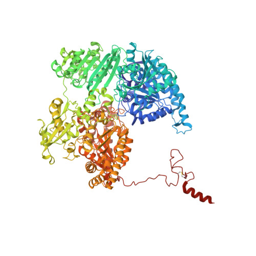
    Pyruvate-ferredoxin oxidoreductases (PFOR) are unique among thiamine pyrophosphate (ThDP)-containing enzymes in giving rise to a rather stable cofactor-based free-radical species upon the decarboxylation of their first substrate, pyruvate. We have obtained snapshots of unreacted and partially reacted (probably as a tetrahedral intermediate) pyruvate-PFOR complexes at different time intervals. We conclude that pyruvate decarboxylation involves very limited substrate-to-product movements but a significant displacement of the thiazolium moiety of ThDP. In this respect, PFOR seems to differ substantially from other ThDP-containing enzymes, such as transketolase and pyruvate decarboxylase. In addition, exposure of PFOR to oxygen in the presence of pyruvate results in significant inhibition of catalytic activity, both in solution and in the crystals. Examination of the crystal structure of inhibited PFOR suggests that the loss of activity results from oxime formation at the 4' amino substituent of the pyrimidine moiety of ThDP.


  • Organizational Affiliation
    • Laboratoire de Cristallographie et Cristallogenèse des Protéines, Institut de Biologie Structurale Jean-Pierre Ebel, CEA, UJF, CNRS, 41 rue Jules Horowitz, 38027 Grenoble Cedex 1, France.

Macromolecule Content 

  • Total Structure Weight: 270.67 kDa 
  • Atom Count: 20,782 
  • Modeled Residue Count: 2,461 
  • Deposited Residue Count: 2,462 
  • Unique protein chains: 1

Macromolecules

Find similar proteins by:|  3D Structure
Entity ID: 1
MoleculeChains  Sequence LengthOrganismDetailsImage
PYRUVATE-FERREDOXIN OXIDOREDUCTASE
A, B
1,231Desulfocurvibacter africanusMutation(s): 0 
EC: 1.2.7.1
UniProt
Find proteins for P94692 (Desulfocurvibacter africanus)
Explore P94692 
Go to UniProtKB:  P94692
Entity Groups
Sequence Clusters30% Identity50% Identity70% Identity90% Identity95% Identity100% Identity
UniProt GroupP94692
Sequence Annotations
Expand
Reference Sequence

Small Molecules

Ligands 5 Unique
IDChains Name / Formula / InChI Key2D Diagram3D Interactions
HTL

Query on HTL


Download:Ideal Coordinates CCD File
F [auth A],
M [auth B]
2-ACETYL-THIAMINE DIPHOSPHATE
C14 H21 N4 O8 P2 S
GYRGKLZCJRVYRV-UHFFFAOYSA-O
SF4

Query on SF4


Download:Ideal Coordinates CCD File
C [auth A]
D [auth A]
E [auth A]
J [auth B]
K [auth B]
C [auth A],
D [auth A],
E [auth A],
J [auth B],
K [auth B],
L [auth B]
IRON/SULFUR CLUSTER
Fe4 S4
LJBDFODJNLIPKO-UHFFFAOYSA-N
CO2

Query on CO2


Download:Ideal Coordinates CCD File
I [auth A],
P [auth B]
CARBON DIOXIDE
C O2
CURLTUGMZLYLDI-UHFFFAOYSA-N
CA

Query on CA


Download:Ideal Coordinates CCD File
H [auth A],
O [auth B]
CALCIUM ION
Ca
BHPQYMZQTOCNFJ-UHFFFAOYSA-N
MG

Query on MG


Download:Ideal Coordinates CCD File
G [auth A],
N [auth B]
MAGNESIUM ION
Mg
JLVVSXFLKOJNIY-UHFFFAOYSA-N

Experimental Data & Validation

Experimental Data

  • Method: X-RAY DIFFRACTION
  • Resolution: 1.93 Å
  • R-Value Free:  0.228 (Depositor), 0.181 (DCC) 
  • R-Value Work:  0.186 (Depositor), 0.186 (DCC) 
  • R-Value Observed: 0.186 (Depositor) 
Space Group: P 21 21 21
Unit Cell:
Length ( Å )Angle ( ˚ )
a = 86.081α = 90
b = 145.639β = 90
c = 211.619γ = 90
Software Package:
Software NamePurpose
CNSrefinement
XDSdata reduction
XSCALEdata scaling
CNSphasing

Structure Validation

View Full Validation Report


Ligand Structure Quality Assessment 


Entry History 

Deposition Data

Revision History  (Full details and data files)

  • Version 1.0: 2006-02-15
    Type: Initial release
  • Version 1.1: 2011-05-07
    Changes: Version format compliance
  • Version 1.2: 2011-07-13
    Changes: Version format compliance
  • Version 1.3: 2023-12-13
    Changes: Data collection, Database references, Derived calculations, Other, Refinement description, Structure summary
  • Version 1.4: 2024-11-13
    Changes: Structure summary