2CN8

Crystal structure of human Chk2 in complex with debromohymenialdisine


Experimental Data Snapshot

  • Method: X-RAY DIFFRACTION
  • Resolution: 2.70 Å
  • R-Value Free: 
    0.271 (Depositor), 0.260 (DCC) 
  • R-Value Work: 
    0.204 (Depositor), 0.200 (DCC) 
  • R-Value Observed: 
    0.207 (Depositor) 

Starting Model: experimental
View more details

wwPDB Validation   3D Report Full Report


Ligand Structure Quality Assessment 

Created with Raphaël 2.3.0Worse 01 BetterLigand structure goodness of fit to experimental dataBest fitted DBQClick on this verticalbar to view details

This is version 1.2 of the entry. See complete history


Literature

Trans-Activation of the DNA-Damage Signalling Protein Kinase Chk2 by T-Loop Exchange

Oliver, A.W.Paul, A.Boxall, K.J.Barrie, S.E.Aherne, G.W.Garrett, M.D.Mittnacht, S.Pearl, L.H.

(2006) EMBO J 25: 3179

  • DOI: https://doi.org/10.1038/sj.emboj.7601209
  • Primary Citation of Related Structures:  
    2CN5, 2CN8

  • PubMed Abstract: 

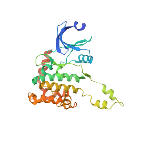
    The protein kinase Chk2 (checkpoint kinase 2) is a major effector of the replication checkpoint. Chk2 activation is initiated by phosphorylation of Thr68, in the serine-glutamine/threonine-glutamine cluster domain (SCD), by ATM. The phosphorylated SCD-segment binds to the FHA domain of a second Chk2 molecule, promoting dimerisation of the protein and triggering phosphorylation of the activation segment/T-loop in the kinase domain. We have now determined the structure of the kinase domain of human Chk2 in complexes with ADP and a small-molecule inhibitor debromohymenialdisine. The structure reveals a remarkable dimeric arrangement in which T-loops are exchanged between protomers, to form an active kinase conformation in trans. Biochemical data suggest that this dimer is the biologically active state promoted by ATM-phosphorylation, and also suggests a mechanism for dimerisation-driven activation of Chk2 by trans-phosphorylation.


  • Organizational Affiliation

    Section of Structural Biology, Cancer Research UK DNA Repair Enzymes Group, The Institute of Cancer Research, Chelsea, London, UK. antony.oliver@icr.ac.uk


Macromolecules
Find similar proteins by:  (by identity cutoff)  |  3D Structure
Entity ID: 1
MoleculeChains Sequence LengthOrganismDetailsImage
SERINE/THREONINE-PROTEIN KINASE CHK2329Homo sapiensMutation(s): 0 
EC: 2.7.11.1
UniProt & NIH Common Fund Data Resources
Find proteins for O96017 (Homo sapiens)
Explore O96017 
Go to UniProtKB:  O96017
PHAROS:  O96017
GTEx:  ENSG00000183765 
Entity Groups  
Sequence Clusters30% Identity50% Identity70% Identity90% Identity95% Identity100% Identity
UniProt GroupO96017
Sequence Annotations
Expand
  • Reference Sequence
Small Molecules
Binding Affinity Annotations 
IDSourceBinding Affinity
DBQ BindingDB:  2CN8 IC50: min: 180, max: 3500 (nM) from 3 assay(s)
Experimental Data & Validation

Experimental Data

  • Method: X-RAY DIFFRACTION
  • Resolution: 2.70 Å
  • R-Value Free:  0.271 (Depositor), 0.260 (DCC) 
  • R-Value Work:  0.204 (Depositor), 0.200 (DCC) 
  • R-Value Observed: 0.207 (Depositor) 
Space Group: P 32 2 1
Unit Cell:
Length ( Å )Angle ( ˚ )
a = 88.65α = 90
b = 88.65β = 90
c = 88.897γ = 120
Software Package:
Software NamePurpose
REFMACrefinement
MOSFLMdata reduction
SCALAdata scaling
PHASERphasing

Structure Validation

View Full Validation Report


Ligand Structure Quality Assessment 

Created with Raphaël 2.3.0Worse 01 BetterLigand structure goodness of fit to experimental dataBest fitted DBQClick on this verticalbar to view details

Entry History 

Deposition Data

Revision History  (Full details and data files)

  • Version 1.0: 2006-06-28
    Type: Initial release
  • Version 1.1: 2011-07-13
    Changes: Advisory, Version format compliance
  • Version 1.2: 2023-12-13
    Changes: Data collection, Database references, Derived calculations, Other, Refinement description