Structural determinants for branched-chain aminotransferase isozyme-specific inhibition by the anticonvulsant drug gabapentin
Goto, M., Miyahara, I., Hirotsu, K., Conway, M., Yennawar, N., Islam, M.M., Hutson, S.M.(2005) J Biological Chem 280: 37246-37256
- PubMed: 16141215
- DOI: https://doi.org/10.1074/jbc.M506486200
- Primary Citation of Related Structures:
2A1H, 2COG, 2COI, 2COJ - PubMed Abstract:
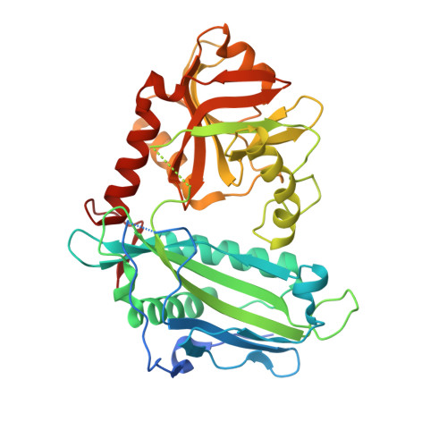
This study presents the first three-dimensional structures of human cytosolic branched-chain aminotransferase (hBCATc) isozyme complexed with the neuroactive drug gabapentin, the hBCATc Michaelis complex with the substrate analog, 4-methylvalerate, and the mitochondrial isozyme (hBCATm) complexed with gabapentin. The branched-chain aminotransferases (BCAT) reversibly catalyze transamination of the essential branched-chain amino acids (leucine, isoleucine, valine) to alpha-ketoglutarate to form the respective branched-chain alpha-keto acids and glutamate. The cytosolic isozyme is the predominant BCAT found in the nervous system, and only hBCATc is inhibited by gabapentin. Pre-steady state kinetics show that 1.3 mm gabapentin can completely inhibit the binding of leucine to reduced hBCATc, whereas 65.4 mm gabapentin is required to inhibit leucine binding to hBCATm. Structural analysis shows that the bulky gabapentin is enclosed in the active-site cavity by the shift of a flexible loop that enlarges the active-site cavity. The specificity of gabapentin for the cytosolic isozyme is ascribed at least in part to the location of the interdomain loop and the relative orientation between the small and large domain which is different from these relationships in the mitochondrial isozyme. Both isozymes contain a CXXC center and form a disulfide bond under oxidizing conditions. The structure of reduced hBCATc was obtained by soaking the oxidized hBCATc crystals with dithiothreitol. The close similarity in active-site structures between cytosolic enzyme complexes in the oxidized and reduced states is consistent with the small effect of oxidation on pre-steady state kinetics of the hBCATc first half-reaction. However, these kinetic data do not explain the inactivation of hBCATm by oxidation of the CXXC center. The structural data suggest that there is a larger effect of oxidation on the interdomain loop and residues surrounding the CXXC center in hBCATm than in hBCATc.
- Department of Chemistry, Graduate School of Science, Osaka City University, Osaka 558-8585, Japan.
Organizational Affiliation:


