2CZY | pdb_00002czy

Solution structure of the NRSF/REST-mSin3B PAH1 complex


Experimental Data Snapshot

  • Method: SOLUTION NMR
  • Conformers Calculated: 200 
  • Conformers Submitted: 20 
  • Selection Criteria: structures with the lowest energy 

wwPDB Validation 3D Report Full Report

Validation slider image for 2CZY

This is version 1.4 of the entry. See complete history

Literature

The Neural Repressor NRSF/REST Binds the PAH1 Domain of the Sin3 Corepressor by Using its Distinct Short Hydrophobic Helix

Nomura, M.Uda-Tochio, H.Murai, K.Mori, N.Nishimura, Y.

(2005) J Mol Biology 354: 903-915

  • DOI: https://doi.org/10.1016/j.jmb.2005.10.008
  • Primary Citation Related Structures: 
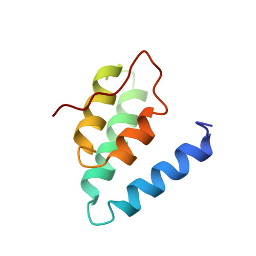
    2CZY

  • PubMed Abstract: 

    In non-neuronal cells and neuronal progenitors, many neuron-specific genes are repressed by a neural restrictive silencer factor (NRSF)/repressor element 1 silencing transcription factor (REST), which is an essential transcriptional repressor recruiting the Sin3-HDAC complex. Sin3 contains four paired amphipathic helix (PAH) domains, PAH1, PAH2, PAH3 and PAH4. A specific target repressor for Sin3 is likely to bind to one of them independently. So far, only the tertiary structures of PAH2 domain complexes, when bound to the Sin3-interacting domains of Mad1 and HBP1, have been determined. Here, we reveal that the N-terminal repressor domain of NRSF/REST binds to the PAH1 domain of mSin3B, and determine the structure of the PAH1 domain associated with the NRSF/REST minimal repressor domain. Compared to the PAH2 structure, PAH1 holds a rather globular four-helix bundle structure with a semi-ordered C-terminal tail. In contrast to the amphipathic alpha-helix of Mad1 or HBP1 bound to PAH2, the short hydrophobic alpha-helix of NRSF/REST is captured in the cleft of PAH1. A nuclear hormone receptor corepressor, N-CoR has been found to bind to the PAH1 domain with a lower affinity than NRSF/REST by using its C-terminal region, which contains fewer hydrophobic amino acid residues than the NRSF/REST helix. For strong binding to a repressor, PAH1 seems to require a short alpha-helix consisting of mostly hydrophobic amino acid residues within the repressor. Each of the four PAH domains of Sin3 seems to interact with a characteristic helix of a specific repressor; PAH1 needs a mostly hydrophobic helix and PAH2 needs an amphipathic helix in each target repressor.


  • Organizational Affiliation
    • Graduate School of Supramolecular Biology Yokohama City University, 1-7-29 Suehiro-cho, Tsurumi-ku, Yokohama 230-0045, Japan.

Macromolecule Content 

  • Total Structure Weight: 10.35 kDa 
  • Atom Count: 731 
  • Modeled Residue Count: 92 
  • Deposited Residue Count: 92 
  • Unique protein chains: 2

Macromolecules

Find similar proteins by:|  3D Structure
Entity ID: 1
MoleculeChains  Sequence LengthOrganismDetailsImage
Paired amphipathic helix protein Sin3b77Mus musculusMutation(s): 0 
Gene Names: Sin3B
UniProt
Find proteins for Q62141 (Mus musculus)
Explore Q62141 
Go to UniProtKB:  Q62141
Entity Groups
Sequence Clusters30% Identity50% Identity70% Identity90% Identity95% Identity100% Identity
UniProt GroupQ62141
Sequence Annotations
Expand
Reference Sequence
Find similar proteins by:  Sequence   |   3D Structure  
Entity ID: 2
MoleculeChains  Sequence LengthOrganismDetailsImage
transcription factor REST (version 3)15N/AMutation(s): 0 
UniProt & NIH Common Fund Data Resources
Find proteins for Q13127 (Homo sapiens)
Explore Q13127 
Go to UniProtKB:  Q13127
PHAROS:  Q13127
GTEx:  ENSG00000084093 
Entity Groups
Sequence Clusters30% Identity50% Identity70% Identity90% Identity95% Identity100% Identity
UniProt GroupQ13127
Sequence Annotations
Expand
Reference Sequence

Experimental Data & Validation

Experimental Data

  • Method: SOLUTION NMR
  • Conformers Calculated: 200 
  • Conformers Submitted: 20 
  • Selection Criteria: structures with the lowest energy 

Structure Validation

View Full Validation Report


Entry History 

Deposition Data

Revision History  (Full details and data files)

  • Version 1.0: 2005-12-20
    Type: Initial release
  • Version 1.1: 2007-10-21
    Changes: Version format compliance
  • Version 1.2: 2011-07-13
    Changes: Version format compliance
  • Version 1.3: 2022-03-09
    Changes: Data collection, Database references, Derived calculations
  • Version 1.4: 2024-05-29
    Changes: Data collection