2FFH | pdb_00002ffh

THE SIGNAL SEQUENCE BINDING PROTEIN FFH FROM THERMUS AQUATICUS


Experimental Data Snapshot

  • Method: X-RAY DIFFRACTION
  • Resolution: 3.20 Å
  • R-Value Free: 
    0.296 (Depositor) 
  • R-Value Work: 
    0.257 (Depositor) 

Starting Model: experimental
View more details

wwPDB Validation 3D Report Full Report

Validation slider image for 2FFH

This is version 1.3 of the entry. See complete history

Literature

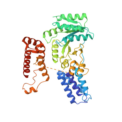
Crystal structure of the signal sequence binding subunit of the signal recognition particle.

Keenan, R.J.Freymann, D.M.Walter, P.Stroud, R.M.

(1998) Cell 94: 181-191

  • DOI: https://doi.org/10.1016/s0092-8674(00)81418-x
  • Primary Citation Related Structures: 
    2FFH

  • PubMed Abstract: 

    The crystal structure of the signal sequence binding subunit of the signal recognition particle (SRP) from Thermus aquaticus reveals a deep groove bounded by a flexible loop and lined with side chains of conserved hydrophobic residues. The groove defines a flexible, hydrophobic environment that is likely to contribute to the structural plasticity necessary for SRP to bind signal sequences of different lengths and amino acid sequence. The structure also reveals a helix-turn-helix motif containing an arginine-rich alpha helix that is required for binding to SRP RNA and is implicated in forming the core of an extended RNA binding surface.


  • Organizational Affiliation
    • Department of Biochemistry and Biophysics, School of Medicine, University of California, San Francisco 94143-0448, USA.

Macromolecule Content 

  • Total Structure Weight: 142.52 kDa 
  • Atom Count: 9,484 
  • Modeled Residue Count: 1,221 
  • Deposited Residue Count: 1,275 
  • Unique protein chains: 1

Macromolecules

Find similar proteins by:|  3D Structure
Entity ID: 1
MoleculeChains  Sequence LengthOrganismDetailsImage
PROTEIN (FFH)
A, B, C
425Thermus aquaticusMutation(s): 0 
EC: 3.6.5.4
UniProt
Find proteins for O07347 (Thermus aquaticus)
Explore O07347 
Go to UniProtKB:  O07347
Entity Groups
Sequence Clusters30% Identity50% Identity70% Identity90% Identity95% Identity100% Identity
UniProt GroupO07347
Sequence Annotations
Expand
Reference Sequence

Small Molecules

Ligands 2 Unique
IDChains Name / Formula / InChI Key2D Diagram3D Interactions
CD

Query on CD


Download:Ideal Coordinates CCD File
D [auth A]
E [auth A]
F [auth A]
G [auth A]
H [auth A]
D [auth A],
E [auth A],
F [auth A],
G [auth A],
H [auth A],
J [auth B],
K [auth B],
L [auth B],
M [auth B],
O [auth C],
P [auth C],
Q [auth C],
R [auth C],
S [auth C],
T [auth C],
U [auth C]
CADMIUM ION
Cd
WLZRMCYVCSSEQC-UHFFFAOYSA-N
SO4

Query on SO4


Download:Ideal Coordinates CCD File
I [auth A],
N [auth B],
V [auth C]
SULFATE ION
O4 S
QAOWNCQODCNURD-UHFFFAOYSA-L

Experimental Data & Validation

Experimental Data

  • Method: X-RAY DIFFRACTION
  • Resolution: 3.20 Å
  • R-Value Free:  0.296 (Depositor) 
  • R-Value Work:  0.257 (Depositor) 
Space Group: H 3 2
Unit Cell:
Length ( Å )Angle ( ˚ )
a = 195.05α = 90
b = 195.05β = 90
c = 335.72γ = 120
Software Package:
Software NamePurpose
CCP4model building
RAVEmodel building
CNSrefinement
DENZOdata reduction
SCALEPACKdata scaling
CCP4phasing
RAVEphasing

Structure Validation

View Full Validation Report


Entry History 

Deposition Data

Revision History  (Full details and data files)

  • Version 1.0: 1999-07-16
    Type: Initial release
  • Version 1.1: 2008-04-27
    Changes: Version format compliance
  • Version 1.2: 2011-07-13
    Changes: Version format compliance
  • Version 1.3: 2023-08-30
    Changes: Data collection, Database references, Derived calculations, Refinement description