2FIR | pdb_00002fir

Crystal structure of DFPR-VIIa/sTF


Experimental Data Snapshot

  • Method: X-RAY DIFFRACTION
  • Resolution: 2.00 Å
  • R-Value Free: 
    0.281 (Depositor), 0.265 (DCC) 
  • R-Value Work: 
    0.207 (Depositor), 0.199 (DCC) 

wwPDB Validation 3D Report Full Report

Validation slider image for 2FIR

Ligand Structure Quality Assessment 


This is version 1.5 of the entry. See complete history

Literature

High Resolution Structures of p-Aminobenzamidine- and Benzamidine-VIIa/Soluble Tissue Factor: Unpredicted conformation of the 192-193 peptide bond and mapping of Ca2+, Mg2+, Na+ and Zn2+ sites in factor VIIa

Bajaj, S.P.Schmidt, A.E.Agah, S.Bajaj, M.S.Padmanabhan, K.

(2006) J Biological Chem 281: 24873-24888

  • DOI: https://doi.org/10.1074/jbc.M509971200
  • Primary Citation Related Structures: 
    2A2Q, 2AER, 2FIR

  • PubMed Abstract: 

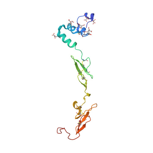
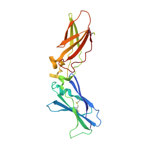
    Factor VIIa (FVIIa) consists of a gamma-carboxyglutamic acid (Gla) domain, two epidermal growth factor-like domains, and a protease domain. FVIIa binds seven Ca(2+) ions in the Gla, one in the EGF1, and one in the protease domain. However, blood contains both Ca(2+) and Mg(2+), and the Ca(2+) sites in FVIIa that could be specifically occupied by Mg(2+) are unknown. Furthermore, FVIIa contains a Na(+) and two Zn(2+) sites, but ligands for these cations are undefined. We obtained p-aminobenzamidine-VIIa/soluble tissue factor (sTF) crystals under conditions containing Ca(2+), Mg(2+), Na(+), and Zn(2+). The crystal diffracted to 1.8A resolution, and the final structure has an R-factor of 19.8%. In this structure, the Gla domain has four Ca(2+) and three bound Mg(2+). The EGF1 domain contains one Ca(2+) site, and the protease domain contains one Ca(2+), one Na(+), and two Zn(2+) sites. (45)Ca(2+) binding in the presence/absence of Mg(2+) to FVIIa, Gla-domainless FVIIa, and prothrombin fragment 1 supports the crystal data. Furthermore, unlike in other serine proteases, the amide N of Gly(193) in FVIIa points away from the oxyanion hole in this structure. Importantly, the oxyanion hole is also absent in the benzamidine-FVIIa/sTF structure at 1.87A resolution. However, soaking benzamidine-FVIIa/sTF crystals with d-Phe-Pro-Arg-chloromethyl ketone results in benzamidine displacement, d-Phe-Pro-Arg incorporation, and oxyanion hole formation by a flip of the 192-193 peptide bond in FVIIa. Thus, it is the substrate and not the TF binding that induces oxyanion hole formation and functional active site geometry in FVIIa. Absence of oxyanion hole is unusual and has biologic implications for FVIIa macromolecular substrate specificity and catalysis.


  • Organizational Affiliation
    • Protein Science Laboratory, UCLA/Orthopaedic Hospital, Department of Orthopaedic Surgery and Molecular Biology Institute, UCLA, Los Angeles, California 90095, USA.

Macromolecule Content 

  • Total Structure Weight: 69.13 kDa 
  • Atom Count: 5,352 
  • Modeled Residue Count: 587 
  • Deposited Residue Count: 601 
  • Unique protein chains: 3

Macromolecules

Find similar proteins by:|  3D Structure
Entity ID: 1
MoleculeChains  Sequence LengthOrganismDetailsImage
Coagulation factor VII Light chainA [auth L]142Homo sapiensMutation(s): 0 
Gene Names: F7
EC: 3.4.21.21
UniProt & NIH Common Fund Data Resources
Find proteins for P08709 (Homo sapiens)
Explore P08709 
Go to UniProtKB:  P08709
PHAROS:  P08709
GTEx:  ENSG00000057593 
Entity Groups
Sequence Clusters30% Identity50% Identity70% Identity90% Identity95% Identity100% Identity
UniProt GroupP08709
Sequence Annotations
Expand
Reference Sequence
Find similar proteins by:|  3D Structure
Entity ID: 2
MoleculeChains  Sequence LengthOrganismDetailsImage
Coagulation factor VII Heavy Chain (EC 3.4.21.21)B [auth H]254Homo sapiensMutation(s): 0 
Gene Names: F7
EC: 3.4.21.21
UniProt & NIH Common Fund Data Resources
Find proteins for P08709 (Homo sapiens)
Explore P08709 
Go to UniProtKB:  P08709
PHAROS:  P08709
GTEx:  ENSG00000057593 
Entity Groups
Sequence Clusters30% Identity50% Identity70% Identity90% Identity95% Identity100% Identity
UniProt GroupP08709
Sequence Annotations
Expand
Reference Sequence
Find similar proteins by:|  3D Structure
Entity ID: 3
MoleculeChains  Sequence LengthOrganismDetailsImage
Tissue factorC [auth T]205Homo sapiensMutation(s): 0 
Gene Names: F3
UniProt & NIH Common Fund Data Resources
Find proteins for P13726 (Homo sapiens)
Explore P13726 
Go to UniProtKB:  P13726
PHAROS:  P13726
GTEx:  ENSG00000117525 
Entity Groups
Sequence Clusters30% Identity50% Identity70% Identity90% Identity95% Identity100% Identity
UniProt GroupP13726
Sequence Annotations
Expand
Reference Sequence

Small Molecules

Ligands 8 Unique
IDChains Name / Formula / InChI Key2D Diagram3D Interactions
0G7

Query on 0G7


Download:Ideal Coordinates CCD File
N [auth H]D-phenylalanyl-N-[(3S)-6-carbamimidamido-1-chloro-2-oxohexan-3-yl]-L-prolinamide
C21 H31 Cl N6 O3
KWPACVJPAFGBEQ-IKGGRYGDSA-N
GLC

Query on GLC


Download:Ideal Coordinates CCD File
D [auth L]alpha-D-glucopyranose
C6 H12 O6
WQZGKKKJIJFFOK-DVKNGEFBSA-N
FUC

Query on FUC


Download:Ideal Coordinates CCD File
E [auth L]alpha-L-fucopyranose
C6 H12 O5
SHZGCJCMOBCMKK-SXUWKVJYSA-N
ZN

Query on ZN


Download:Ideal Coordinates CCD File
Q [auth H],
R [auth H]
ZINC ION
Zn
PTFCDOFLOPIGGS-UHFFFAOYSA-N
CA

Query on CA


Download:Ideal Coordinates CCD File
G [auth L]
H [auth L]
J [auth L]
K [auth L]
M [auth L]
G [auth L],
H [auth L],
J [auth L],
K [auth L],
M [auth L],
O [auth H]
CALCIUM ION
Ca
BHPQYMZQTOCNFJ-UHFFFAOYSA-N
CL

Query on CL


Download:Ideal Coordinates CCD File
S [auth H],
T [auth H],
U [auth T]
CHLORIDE ION
Cl
VEXZGXHMUGYJMC-UHFFFAOYSA-M
MG

Query on MG


Download:Ideal Coordinates CCD File
F [auth L],
I [auth L],
L
MAGNESIUM ION
Mg
JLVVSXFLKOJNIY-UHFFFAOYSA-N
NA

Query on NA


Download:Ideal Coordinates CCD File
P [auth H]SODIUM ION
Na
FKNQFGJONOIPTF-UHFFFAOYSA-N
Modified Residues  1 Unique
IDChains TypeFormula2D DiagramParent
CGU
Query on CGU
A [auth L]L-PEPTIDE LINKINGC6 H9 N O6GLU

Biologically Interesting Molecules (External Reference) 

1 Unique

Experimental Data & Validation

Experimental Data

  • Method: X-RAY DIFFRACTION
  • Resolution: 2.00 Å
  • R-Value Free:  0.281 (Depositor), 0.265 (DCC) 
  • R-Value Work:  0.207 (Depositor), 0.199 (DCC) 
Space Group: P 21 21 21
Unit Cell:
Length ( Å )Angle ( ˚ )
a = 70.02α = 90
b = 80.98β = 90
c = 126.33γ = 90
Software Package:
Software NamePurpose
X-PLORmodel building
X-PLORrefinement
HKL-2000data reduction
SCALEPACKdata scaling
X-PLORphasing

Structure Validation

View Full Validation Report


Ligand Structure Quality Assessment 


Entry History 

Deposition Data

Revision History  (Full details and data files)

  • Version 1.0: 2006-07-11
    Type: Initial release
  • Version 1.1: 2008-05-01
    Changes: Version format compliance
  • Version 1.2: 2011-07-13
    Changes: Atomic model, Database references, Derived calculations, Non-polymer description, Structure summary, Version format compliance
  • Version 1.3: 2013-02-27
    Changes: Other
  • Version 1.4: 2020-07-29
    Type: Remediation
    Reason: Carbohydrate remediation
    Changes: Data collection, Derived calculations, Structure summary
  • Version 1.5: 2024-12-25
    Changes: Advisory, Data collection, Database references, Derived calculations, Structure summary