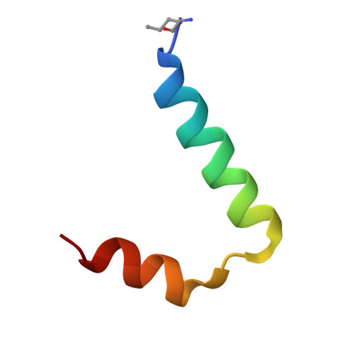
Diabetes mellitus due to misfolding of a beta-cell transcription factor: stereospecific frustration of a Schellman motif in HNF-1alpha.
Narayana, N., Phillips, N.B., Hua, Q.X., Jia, W., Weiss, M.A.(2006) J Mol Biology 362: 414-429
- PubMed: 16930618
- DOI: https://doi.org/10.1016/j.jmb.2006.06.086
- Primary Citation of Related Structures:
2GYP - PubMed Abstract:
Maturity-onset diabetes of the young (MODY3), a monogenic form of type II diabetes mellitus, results most commonly from mutations in hepatocyte nuclear factor 1alpha (HNF-1alpha). Diabetes-associated mutation G20R perturbs the dimerization domain of HNF-1alpha, an intertwined four-helix bundle. In the wild-type structure G20 participates in a Schellman motif to cap an alpha-helix; its dihedral angles lie in the right side of the Ramachandran plot (alpha(L) region; phi 97 degrees). Substitutions G20R and G20A lead to dimeric molten globules of low stability, suggesting that the impaired function of the diabetes-associated transcription factor is due in large part to a main-chain perturbation rather than to specific features of the Arg side-chain. This hypothesis is supported by the enhanced stability of non-standard analogues containing D-Ala or D-Ser at position 20. The crystal structure of the D-Ala20 analogue, determined to a resolution of 1.4 A, is essentially identical to the wild-type structure in the same crystal form. The mean root-mean-square deviation between equivalent C(alpha) atoms (residues 5-28) is 0.3 A; (phi, psi) angles of D-Ala20 are the same as those of G20 in the wild-type structure. Whereas the side-chain of A20 or R20 would be expected to clash with the preceding carbonyl oxygen (thus accounting for its frustrated energy landscape), the side-chain of D-Ala20 projects into solvent without perturbation of the Schellman motif. Calorimetric studies indicate that the increased stability of the D-Ala20 analogue (DeltaDeltaG(u) 1.5 kcal/mol) is entropic in origin, consistent with a conformational bias toward native-like conformations in the unfolded state. Studies of multiple substitutions at G20 and neighboring positions highlight the essential contributions of a glycine-specific tight turn and adjoining inter-subunit side-chain hydrogen bonds to the stability and architectural specificity of the intertwined dimer. Comparison of L- and D amino acid substitutions thus provides an example of the stereospecific control of an energy landscape by a helix-capping residue.
- Department of Biochemistry, Case Western Reserve University, 10900 Euclid Avenue, Cleveland, OH 44106-4935, USA.
Organizational Affiliation:


