2HBH | pdb_00002hbh

Crystal structure of Vitamin D nuclear receptor ligand binding domain bound to a locked side-chain analog of calcitriol and SRC-1 peptide


Experimental Data Snapshot

  • Method: X-RAY DIFFRACTION
  • Resolution: 2.65 Å
  • R-Value Free: 
    0.261 (Depositor), 0.254 (DCC) 
  • R-Value Work: 
    0.203 (Depositor), 0.190 (DCC) 
  • R-Value Observed: 
    0.206 (Depositor) 

Starting Model: experimental
View more details

wwPDB Validation 3D Report Full Report

Validation slider image for 2HBH

Ligand Structure Quality Assessment 


This is version 1.4 of the entry. See complete history

Literature

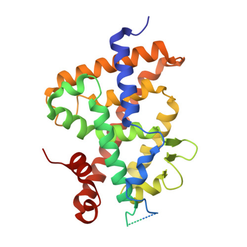
Crystal structure of the vitamin D nuclear receptor ligand binding domain in complex with a locked side chain analog of calcitriol

Rochel, N.Hourai, S.Perez-Garcia, X.Rumbo, A.Mourino, A.Moras, D.

(2007) Arch Biochem Biophys 460: 172-176

  • DOI: https://doi.org/10.1016/j.abb.2007.01.031
  • Primary Citation Related Structures: 
    2HBH

  • PubMed Abstract: 

    The crystal structures of vitamin D nuclear receptor (VDR) have revealed that all compounds are anchored by the same residues to the ligand binding pocket (LBP). Based on this observation, a synthetic analog with a locked side chain (21-nor-calcitriol-20(22),23-diyne) has been synthesized in order to gain in entropy energy with a predefined active side chain conformation. The crystal structure of VDR LBD bound to this locked side chain analogue while confirming the docking provides a structural basis for the activity of this compound.


  • Organizational Affiliation
    • Institut de Génétique et de Biologie Moléculaire et Cellulaire, Département de Biologie et de Génomique Structurales, Illkirch F-67400, France.

Macromolecule Content 

  • Total Structure Weight: 36.23 kDa 
  • Atom Count: 2,109 
  • Modeled Residue Count: 249 
  • Deposited Residue Count: 317 
  • Unique protein chains: 2

Macromolecules

Find similar proteins by:|  3D Structure
Entity ID: 1
MoleculeChains  Sequence LengthOrganismDetailsImage
Vitamin D receptor302Danio rerioMutation(s): 0 
UniProt
Find proteins for Q9PTN2 (Danio rerio)
Explore Q9PTN2 
Go to UniProtKB:  Q9PTN2
Entity Groups
Sequence Clusters30% Identity50% Identity70% Identity90% Identity95% Identity100% Identity
UniProt GroupQ9PTN2
Sequence Annotations
Expand
Reference Sequence
Find similar proteins by:  Sequence   |   3D Structure  
Entity ID: 2
MoleculeChains  Sequence LengthOrganismDetailsImage
SRC-1 from Nuclear receptor coactivator 115Homo sapiensMutation(s): 0 
EC: 2.3.1.48
UniProt & NIH Common Fund Data Resources
Find proteins for Q15788 (Homo sapiens)
Explore Q15788 
Go to UniProtKB:  Q15788
PHAROS:  Q15788
GTEx:  ENSG00000084676 
Entity Groups
Sequence Clusters30% Identity50% Identity70% Identity90% Identity95% Identity100% Identity
UniProt GroupQ15788
Sequence Annotations
Expand
Reference Sequence

Small Molecules

Ligands 1 Unique
IDChains Name / Formula / InChI Key2D Diagram3D Interactions
XE4

Query on XE4


Download:Ideal Coordinates CCD File
C [auth A]1,3-CYCLOHEXANEDIOL, 4-METHYLENE-5-[(2E)-[(1S,3AS,7AS)-OCTAHYDRO-1-(5-HYDROXY-5-METHYL-1,3-HEXADIYNYL)-7A-METHYL-4H-INDEN-4-YLIDENE]ETHYLIDENE]-, (1R,3S,5Z)
C26 H34 O3
CEEUUHVULXTFGS-BQXVGYHGSA-N

Experimental Data & Validation

Experimental Data

  • Method: X-RAY DIFFRACTION
  • Resolution: 2.65 Å
  • R-Value Free:  0.261 (Depositor), 0.254 (DCC) 
  • R-Value Work:  0.203 (Depositor), 0.190 (DCC) 
  • R-Value Observed: 0.206 (Depositor) 
Space Group: P 65 2 2
Unit Cell:
Length ( Å )Angle ( ˚ )
a = 65.682α = 90
b = 65.682β = 90
c = 265.096γ = 120
Software Package:
Software NamePurpose
CNSrefinement
ADSCdata collection
HKL-2000data scaling
CNSphasing

Structure Validation

View Full Validation Report


Ligand Structure Quality Assessment 


Entry History 

Deposition Data

Revision History  (Full details and data files)

  • Version 1.0: 2007-05-01
    Type: Initial release
  • Version 1.1: 2008-05-01
    Changes: Version format compliance
  • Version 1.2: 2011-07-13
    Changes: Version format compliance
  • Version 1.3: 2021-03-03
    Changes: Database references, Derived calculations, Source and taxonomy
  • Version 1.4: 2023-10-25
    Changes: Data collection, Database references, Refinement description