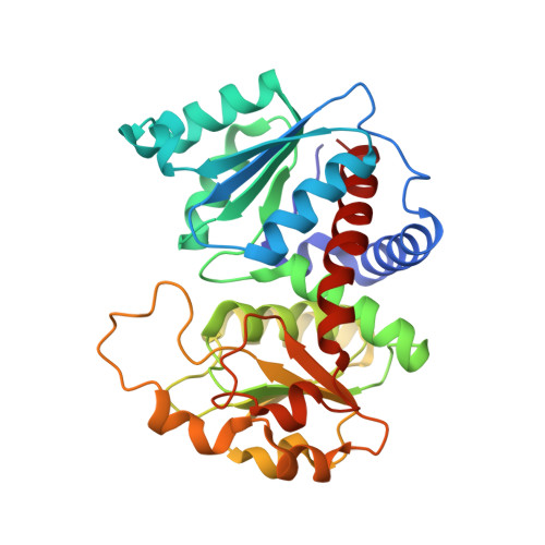
2I6U

Crystal Structure of Ornithine Carbamoyltransferase complexed with Carbamoyl Phosphate and L-Norvaline from Mycobacterium tuberculosis (Rv1656) at 2.2 A


Experimental Data Snapshot

  • Method: X-RAY DIFFRACTION
  • Resolution: 2.20 Å
  • R-Value Free: 0.239 
  • R-Value Work: 0.176 
  • R-Value Observed: 0.179 

Starting Model: experimental
View more details

wwPDB Validation   3D Report Full Report


This is version 1.3 of the entry. See complete history


Literature

The crystal structures of ornithine carbamoyltransferase from Mycobacterium tuberculosis and its ternary complex with carbamoyl phosphate and L-norvaline reveal the enzyme's catalytic mechanism

Sankaranarayanan, R.Cherney, M.M.Cherney, L.T.Garen, C.R.Moradian, F.James, M.N.

(2008) J Mol Biol 375: 1052-1063

  • DOI: https://doi.org/10.1016/j.jmb.2007.11.025
  • Primary Citation of Related Structures:  
    2I6U, 2P2G

  • PubMed Abstract: 

    Mycobacterium tuberculosis ornithine carbamoyltransferase (Mtb OTC) catalyzes the sixth step in arginine biosynthesis; it produces citrulline from carbamoyl phosphate (CP) and ornithine (ORN). Here, we report the crystal structures of Mtb OTC in orthorhombic (form I) and hexagonal (form II) space groups. The molecules in form II are complexed with CP and l-norvaline (NVA); the latter is a competitive inhibitor of OTC. The asymmetric unit in form I contains a pseudo hexamer with 32 point group symmetry. The CP and NVA in form II induce a remarkable conformational change in the 80s and the 240s loops with the displacement of these loops towards the active site. The displacement of these loops is strikingly different from that seen in other OTC structures. In addition, the ligands induce a domain closure of 4.4 degrees in form II. Sequence comparison of active-site residues of Mtb OTC with several other OTCs of known structure reveals that they are virtually identical. The interactions involving the active-site residues of Mtb OTC with CP and NVA and a modeling study of ORN in the form II structure strongly rule out an earlier proposed mechanistic role of Cys264 in catalysis and suggest a possible mechanism for OTC. Our results strongly support the view that ORN with an already deprotonated N(epsilon) atom is the species that binds to the enzyme and that one of the phosphate oxygen atoms of CP is likely to be involved in accepting a proton from the doubly protonated N(epsilon) atom of ORN. We have interpreted this deprotonation as part of the collapse of the transition state of the reaction.


  • Organizational Affiliation

    Group in Protein Structure and Function, Department of Biochemistry, University of Alberta, Edmonton, Alberta, Canada.


Macromolecules
Find similar proteins by:  (by identity cutoff)  |  3D Structure
Entity ID: 1
MoleculeChains Sequence LengthOrganismDetailsImage
Ornithine carbamoyltransferase
A, B, C
307Mycobacterium tuberculosisMutation(s): 0 
Gene Names: argFRv1656MT1694MTCY06H11.21
EC: 2.1.3.3
UniProt
Find proteins for P9WIT9 (Mycobacterium tuberculosis (strain ATCC 25618 / H37Rv))
Explore P9WIT9 
Go to UniProtKB:  P9WIT9
Entity Groups  
Sequence Clusters30% Identity50% Identity70% Identity90% Identity95% Identity100% Identity
UniProt GroupP9WIT9
Sequence Annotations
Expand
  • Reference Sequence
Small Molecules
Ligands 3 Unique
IDChains Name / Formula / InChI Key2D Diagram3D Interactions
CP
Query on CP

Download Ideal Coordinates CCD File 
F [auth A],
I [auth B],
M [auth C]
PHOSPHORIC ACID MONO(FORMAMIDE)ESTER
C H4 N O5 P
FFQKYPRQEYGKAF-UHFFFAOYSA-N
NVA
Query on NVA

Download Ideal Coordinates CCD File 
G [auth A],
J [auth B],
N [auth C]
NORVALINE
C5 H11 N O2
SNDPXSYFESPGGJ-BYPYZUCNSA-N
SO4
Query on SO4

Download Ideal Coordinates CCD File 
D [auth A],
E [auth A],
H [auth B],
K [auth C],
L [auth C]
SULFATE ION
O4 S
QAOWNCQODCNURD-UHFFFAOYSA-L
Experimental Data & Validation

Experimental Data

  • Method: X-RAY DIFFRACTION
  • Resolution: 2.20 Å
  • R-Value Free: 0.239 
  • R-Value Work: 0.176 
  • R-Value Observed: 0.179 
  • Space Group: P 65 2 2
Unit Cell:
Length ( Å )Angle ( ˚ )
a = 99.149α = 90
b = 99.149β = 90
c = 463γ = 120
Software Package:
Software NamePurpose
REFMACrefinement
Blu-Icedata collection
HKL-2000data reduction
SCALEPACKdata scaling
AMoREphasing

Structure Validation

View Full Validation Report


Entry History 

Revision History  (Full details and data files)

  • Version 1.0: 2007-06-26
    Type: Initial release
  • Version 1.1: 2008-04-21
    Changes: Version format compliance
  • Version 1.2: 2011-07-13
    Changes: Version format compliance
  • Version 1.3: 2023-08-30
    Changes: Data collection, Database references, Derived calculations, Refinement description