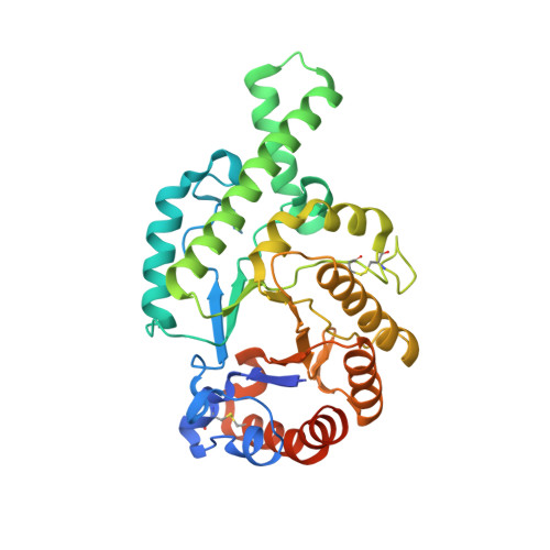
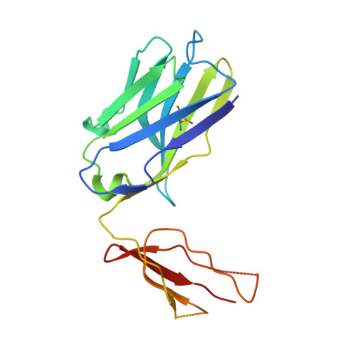
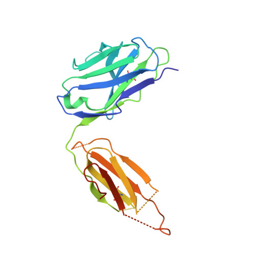
2J88

Hyaluronidase in complex with a monoclonal IgG Fab fragment


Experimental Data Snapshot

  • Method: X-RAY DIFFRACTION
  • Resolution: 2.60 Å
  • R-Value Free: 0.247 
  • R-Value Work: 0.210 
  • R-Value Observed: 0.212 

Starting Models: experimental
View more details

wwPDB Validation   3D Report Full Report


This is version 1.4 of the entry. See complete history


Literature

Identification of a B-Cell Epitope of Hyaluronidase, a Major Bee Venom Allergen, from its Crystal Structure in Complex with a Specific Fab.

Padavattan, S.Schirmer, T.Schmidt, M.Akdis, C.Valenta, R.Mittermann, I.Soldatova, L.Slater, J.Mueller, U.Markovic-Housley, Z.

(2007) J Mol Biol 368: 742

  • DOI: https://doi.org/10.1016/j.jmb.2007.02.036
  • Primary Citation of Related Structures:  
    2J88

  • PubMed Abstract: 

    The major allergens of honeybee venom, hyaluronidase (Hyal) and phospholipase A2, can induce life-threatening IgE-mediated allergic reactions in humans. Although conventional immunotherapy is effective, up to 40% of patients develop allergic side effects including anaphylaxis and thus, there is a need for an improved immunotherapy. A murine monoclonal anti-Hyal IgG1 antibody (mAb 21E11), that competed for Hyal binding with IgEs from sera of bee venom allergic patients, was raised. The fragment of these IgG antibodies which bind to antigen (Fab) was produced and complexed (1:1) with Hyal. The crystal structure determination of Hyal/Fab 21E11 complex (2.6 A) enabled the identification of the Hyal-IgG interface which provides indirect information on the Hyal-IgE interaction (B-cell epitope). The epitope is composed of a linear array of nine residues (Arg138, His141-Arg148) located at the tip of a helix-turn-helix motive which protrudes away from the globular core and fits tightly into the deep surface pocket formed by the residues from the six complementarity determining regions (CDRs) of the Fab. The epitope is continuous and yet its conformation appears to be essential for Ab recognition, since the synthetic 15-mer peptide comprising the entire epitope (Arg138-Glu152) is neither recognized by mAb 21E11 nor by human IgEs. The structure of the complex provides the basis for the rational design of Hyal derivatives with reduced allergenic activity, which could be used in the development of safer allergen-specific immunotherapy.


  • Organizational Affiliation

    Division of Structural Biology, Biozentrum, University of Basel, CH-4056 Basel, Switzerland.


Macromolecules
Find similar proteins by:  (by identity cutoff)  |  3D Structure
Entity ID: 1
MoleculeChains Sequence LengthOrganismDetailsImage
HYALURONONGLUCOSAMINIDASE350Apis melliferaMutation(s): 0 
EC: 3.2.1.35
UniProt
Find proteins for Q08169 (Apis mellifera)
Explore Q08169 
Go to UniProtKB:  Q08169
Entity Groups  
Sequence Clusters30% Identity50% Identity70% Identity90% Identity95% Identity100% Identity
UniProt GroupQ08169
Sequence Annotations
Expand
  • Reference Sequence
Find similar proteins by:  (by identity cutoff)  |  3D Structure
Entity ID: 2
MoleculeChains Sequence LengthOrganismDetailsImage
FABB [auth H]191Mus musculusMutation(s): 0 
Entity Groups  
Sequence Clusters30% Identity50% Identity70% Identity90% Identity95% Identity100% Identity
Sequence Annotations
Expand
  • Reference Sequence
Find similar proteins by:  (by identity cutoff)  |  3D Structure
Entity ID: 3
MoleculeChains Sequence LengthOrganismDetailsImage
FABC [auth L]213Mus musculusMutation(s): 0 
Entity Groups  
Sequence Clusters30% Identity50% Identity70% Identity90% Identity95% Identity100% Identity
Sequence Annotations
Expand
  • Reference Sequence
Experimental Data & Validation

Experimental Data

  • Method: X-RAY DIFFRACTION
  • Resolution: 2.60 Å
  • R-Value Free: 0.247 
  • R-Value Work: 0.210 
  • R-Value Observed: 0.212 
  • Space Group: C 1 2 1
Unit Cell:
Length ( Å )Angle ( ˚ )
a = 151.563α = 90
b = 70.05β = 123.68
c = 138.88γ = 90
Software Package:
Software NamePurpose
REFMACrefinement
MOSFLMdata reduction
SCALAdata scaling
PHASERphasing

Structure Validation

View Full Validation Report


Entry History 

Deposition Data

Revision History  (Full details and data files)

  • Version 1.0: 2007-04-03
    Type: Initial release
  • Version 1.1: 2011-07-13
    Changes: Advisory, Refinement description, Version format compliance
  • Version 1.2: 2014-02-05
    Changes: Database references, Derived calculations, Source and taxonomy
  • Version 1.3: 2023-12-13
    Changes: Data collection, Database references, Other, Refinement description
  • Version 1.4: 2024-10-09
    Changes: Structure summary