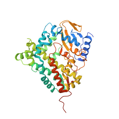
2JJP

Structure of cytochrome P450 EryK in complex with inhibitor ketoconazole (KC)


Experimental Data Snapshot

  • Method: X-RAY DIFFRACTION
  • Resolution: 2.10 Å
  • R-Value Free: 0.262 
  • R-Value Work: 0.195 
  • R-Value Observed: 0.199 

Starting Model: experimental
View more details

wwPDB Validation   3D Report Full Report


Ligand Structure Quality Assessment 


This is version 1.2 of the entry. See complete history


Literature

Azole Drugs Trap Cytochrome P450 Eryk in Alternative Conformational States.

Montemiglio, L.C.Gianni, S.Vallone, B.Savino, C.

(2010) Biochemistry 49: 9199

  • DOI: https://doi.org/10.1021/bi101062v
  • Primary Citation of Related Structures:  
    2JJP, 2XFH

  • PubMed Abstract: 

    EryK is a bacterial cytochrome P450 that catalyzes the last hydroxylation occurring during the biosynthetic pathway of erythromycin A in Streptomyces erythraeus. We report the crystal structures of EryK in complex with two widely used azole inhibitors: ketoconazole and clotrimazole. Both of these ligands use their imidazole moiety to coordinate the heme iron of P450s. Nevertheless, because of the different chemical and structural properties of their N1-substituent group, ketoconazole and clotrimazole trap EryK, respectively, in a closed and in an open conformation that resemble the two structures previously described for the ligand-free EryK. Indeed, ligands induce a distortion of the internal helix I that affects the accessibility of the binding pocket by regulating the kink of the external helix G via a network of interactions that involves helix F. The data presented thus constitute an example of how a cytochrome P450 may be selectively trapped in different conformational states by inhibitors.


  • Organizational Affiliation

    Department of Biochemical Sciences, Sapienza University of Rome and CNR Institute of Molecular Biology and Pathology, Piazzale A. Moro 5, Rome, Italy.


Macromolecules
Find similar proteins by:  (by identity cutoff)  |  3D Structure
Entity ID: 1
MoleculeChains Sequence LengthOrganismDetailsImage
CYTOCHROME P450 113A1411Saccharopolyspora erythraeaMutation(s): 2 
EC: 1.14 (PDB Primary Data), 1.14.13.154 (UniProt)
UniProt
Find proteins for P48635 (Saccharopolyspora erythraea (strain ATCC 11635 / DSM 40517 / JCM 4748 / NBRC 13426 / NCIMB 8594 / NRRL 2338))
Explore P48635 
Go to UniProtKB:  P48635
Entity Groups  
Sequence Clusters30% Identity50% Identity70% Identity90% Identity95% Identity100% Identity
UniProt GroupP48635
Sequence Annotations
Expand
  • Reference Sequence
Small Molecules
Ligands 3 Unique
IDChains Name / Formula / InChI Key2D Diagram3D Interactions
HEM
Query on HEM

Download Ideal Coordinates CCD File 
B [auth A]PROTOPORPHYRIN IX CONTAINING FE
C34 H32 Fe N4 O4
KABFMIBPWCXCRK-RGGAHWMASA-L
KLN
Query on KLN

Download Ideal Coordinates CCD File 
C [auth A]1-ACETYL-4-(4-{[(2S,4R)-2-(2,4-DICHLOROPHENYL)-2-(1H-IMIDAZOL-1-YLMETHYL)-1,3-DIOXOLAN-4-YL]METHOXY}PHENYL)PIPERAZINE
C26 H28 Cl2 N4 O4
XMAYWYJOQHXEEK-ZEQKJWHPSA-N
SO4
Query on SO4

Download Ideal Coordinates CCD File 
D [auth A],
E [auth A]
SULFATE ION
O4 S
QAOWNCQODCNURD-UHFFFAOYSA-L
Experimental Data & Validation

Experimental Data

  • Method: X-RAY DIFFRACTION
  • Resolution: 2.10 Å
  • R-Value Free: 0.262 
  • R-Value Work: 0.195 
  • R-Value Observed: 0.199 
  • Space Group: P 1 21 1
Unit Cell:
Length ( Å )Angle ( ˚ )
a = 53.243α = 90
b = 68.038β = 100.71
c = 57.687γ = 90
Software Package:
Software NamePurpose
REFMACrefinement
DENZOdata reduction
SCALEPACKdata scaling
REFMACphasing

Structure Validation

View Full Validation Report


Ligand Structure Quality Assessment 


Entry History 

Deposition Data

Revision History  (Full details and data files)

  • Version 1.0: 2009-07-14
    Type: Initial release
  • Version 1.1: 2012-08-22
    Changes: Database references, Derived calculations, Non-polymer description, Other, Structure summary, Version format compliance
  • Version 1.2: 2023-12-13
    Changes: Data collection, Database references, Derived calculations, Other, Refinement description