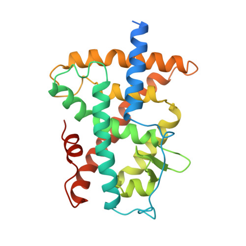
2LBD

LIGAND-BINDING DOMAIN OF THE HUMAN RETINOIC ACID RECEPTOR GAMMA BOUND TO ALL-TRANS RETINOIC ACID


Experimental Data Snapshot

  • Method: X-RAY DIFFRACTION
  • Resolution: 2.06 Å
  • R-Value Free: 0.313 
  • R-Value Work: 0.210 
  • R-Value Observed: 0.210 

wwPDB Validation   3D Report Full Report


Ligand Structure Quality Assessment 


This is version 1.3 of the entry. See complete history


Literature

Crystal structure of the RAR-gamma ligand-binding domain bound to all-trans retinoic acid.

Renaud, J.P.Rochel, N.Ruff, M.Vivat, V.Chambon, P.Gronemeyer, H.Moras, D.

(1995) Nature 378: 681-689

  • DOI: https://doi.org/10.1038/378681a0
  • Primary Citation of Related Structures:  
    2LBD

  • PubMed Abstract: 

    The 2.0-A crystal structure of the ligand-binding domain (LBD) of the human retinoic acid receptor (RAR)-gamma bound to all-trans retinoic acid reveals the ligand-binding interactions and suggests an electrostatic guidance mechanism. The overall fold is similar to that of the human RXR-alpha apo-LBD, except for the carboxy-terminal part which folds back towards the LBD core, contributing to the hydrophobic ligand pocket and 'sealing' its entry site. We propose a 'mouse trap' mechanism whereby a ligand-induced conformational transition repositions the amphipathic alpha-helix of the AF-2 activating domain and forms a transcriptionally active receptor.


  • Organizational Affiliation

    Institut de Génétique et de Biologie Moléculaire et Cellulaire, CNRS/INSERM/ULP/Collège de France, Strasbourg, France.


Macromolecules
Find similar proteins by:  (by identity cutoff)  |  3D Structure
Entity ID: 1
MoleculeChains Sequence LengthOrganismDetailsImage
RETINOIC ACID RECEPTOR GAMMA267Homo sapiensMutation(s): 0 
Gene Names: HUMAN RAR GAMMA A CDNA (NUCLEOTIDES 946 - 1683)
UniProt & NIH Common Fund Data Resources
Find proteins for P13631 (Homo sapiens)
Explore P13631 
Go to UniProtKB:  P13631
PHAROS:  P13631
GTEx:  ENSG00000172819 
Entity Groups  
Sequence Clusters30% Identity50% Identity70% Identity90% Identity95% Identity100% Identity
UniProt GroupP13631
Sequence Annotations
Expand
  • Reference Sequence
Small Molecules
Ligands 1 Unique
IDChains Name / Formula / InChI Key2D Diagram3D Interactions
REA
Query on REA

Download Ideal Coordinates CCD File 
B [auth A]RETINOIC ACID
C20 H28 O2
SHGAZHPCJJPHSC-YCNIQYBTSA-N
Binding Affinity Annotations 
IDSourceBinding Affinity
REA BindingDB:  2LBD IC50: min: 1000, max: 1000 (nM) from 2 assay(s)
Experimental Data & Validation

Experimental Data

  • Method: X-RAY DIFFRACTION
  • Resolution: 2.06 Å
  • R-Value Free: 0.313 
  • R-Value Work: 0.210 
  • R-Value Observed: 0.210 
  • Space Group: P 41 21 2
Unit Cell:
Length ( Å )Angle ( ˚ )
a = 60.6α = 90
b = 60.6β = 90
c = 155.3γ = 90
Software Package:
Software NamePurpose
X-PLORmodel building
X-PLORrefinement
MARXDSdata reduction
MARSCALEdata scaling
X-PLORphasing

Structure Validation

View Full Validation Report


Ligand Structure Quality Assessment 


Entry History 

Deposition Data

Revision History  (Full details and data files)

  • Version 1.0: 1997-11-12
    Type: Initial release
  • Version 1.1: 2008-03-24
    Changes: Version format compliance
  • Version 1.2: 2011-07-13
    Changes: Version format compliance
  • Version 1.3: 2024-02-21
    Changes: Data collection, Database references, Derived calculations, Other