2MQS | pdb_00002mqs

Transient Collagen Triple Helix Binding to a Key Metalloproteinase in Invasion and Development: Spin Labels to Structure


Experimental Data Snapshot

  • Method: SOLUTION NMR
  • Conformers Calculated: 200 
  • Conformers Submitted: 15 
  • Selection Criteria: structures with the lowest energy 

wwPDB Validation 3D Report Full Report

Validation slider image for 2MQS

This is version 1.2 of the entry. See complete history

Literature

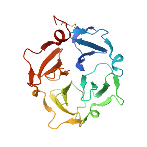
Transient collagen triple helix binding to a key metalloproteinase in invasion and development.

Zhao, Y.Marcink, T.C.Sanganna Gari, R.R.Marsh, B.P.King, G.M.Stawikowska, R.Fields, G.B.Van Doren, S.R.

(2015) Structure 23: 257-269

  • DOI: https://doi.org/10.1016/j.str.2014.11.021
  • Primary Citation Related Structures: 
    2MQS

  • PubMed Abstract: 

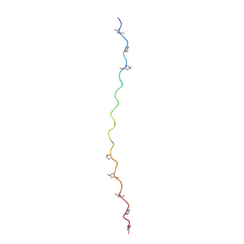
    Skeletal development and invasion by tumor cells depends on proteolysis of collagen by the pericellular metalloproteinase MT1-MMP. Its hemopexin-like (HPX) domain binds to collagen substrates to facilitate their digestion. Spin labeling and paramagnetic nuclear magnetic resonance (NMR) detection have revealed how the HPX domain docks to collagen I-derived triple helix. Mutations impairing triple-helical peptidase activity corroborate the interface. Saturation transfer difference NMR suggests rotational averaging around the longitudinal axis of the triple-helical peptide. Part of the interface emerges as unique and potentially targetable for selective inhibition. The triple helix crosses the junction of blades I and II at a 45° angle to the symmetry axis of the HPX domain, placing the scissile Gly∼Ile bond near the HPX domain and shifted ∼25 Å from MMP-1 complexes. This raises the question of the MT1-MMP catalytic domain folding over the triple helix during catalysis, a possibility accommodated by the flexibility between domains suggested by atomic force microscopy images.


  • Organizational Affiliation
    • Department of Biochemistry, University of Missouri, 117 Schweitzer Hall, Columbia, MO 65211, USA.

Macromolecule Content 

  • Total Structure Weight: 32.74 kDa 
  • Atom Count: 2,315 
  • Modeled Residue Count: 301 
  • Deposited Residue Count: 301 
  • Unique protein chains: 3

Macromolecules

Find similar proteins by:|  3D Structure
Entity ID: 1
MoleculeChains  Sequence LengthOrganismDetailsImage
Matrix metalloproteinase-14196Homo sapiensMutation(s): 0 
Gene Names: MMP14
EC: 3.4.24.80
UniProt & NIH Common Fund Data Resources
Find proteins for P50281 (Homo sapiens)
Explore P50281 
Go to UniProtKB:  P50281
PHAROS:  P50281
GTEx:  ENSG00000157227 
Entity Groups
Sequence Clusters30% Identity50% Identity70% Identity90% Identity95% Identity100% Identity
UniProt GroupP50281
Sequence Annotations
Expand
Reference Sequence
Find similar proteins by:|  3D Structure
Entity ID: 2
MoleculeChains  Sequence LengthOrganismDetailsImage
THP_L_and_M_chain
B, C
36N/AMutation(s): 0 
Find similar proteins by:|  3D Structure
Entity ID: 3
MoleculeChains  Sequence LengthOrganismDetailsImage
THP_T_chain33N/AMutation(s): 0 

Small Molecules

Modified Residues  1 Unique
IDChains TypeFormula2D DiagramParent
HYP
Query on HYP
B, C
L-PEPTIDE LINKINGC5 H9 N O3PRO

Experimental Data & Validation

Experimental Data

  • Method: SOLUTION NMR
  • Conformers Calculated: 200 
  • Conformers Submitted: 15 
  • Selection Criteria: structures with the lowest energy 

Structure Validation

View Full Validation Report


Entry History 

Deposition Data

Revision History  (Full details and data files)

  • Version 1.0: 2015-02-11
    Type: Initial release
  • Version 1.1: 2018-10-10
    Changes: Data collection, Database references, Structure summary
  • Version 1.2: 2023-06-14
    Changes: Data collection, Database references, Derived calculations, Other, Structure summary