2OI5 | pdb_00002oi5

E. coli GlmU- Complex with UDP-GlcNAc and Acetyl-CoA


Experimental Data Snapshot

  • Method: X-RAY DIFFRACTION
  • Resolution: 2.25 Å
  • R-Value Free: 
    0.219 (Depositor), 0.219 (DCC) 
  • R-Value Work: 
    0.176 (Depositor), 0.177 (DCC) 
  • R-Value Observed: 
    0.176 (Depositor) 

Starting Model: experimental
View more details

wwPDB Validation 3D Report Full Report

Validation slider image for 2OI5

Ligand Structure Quality Assessment 


This is version 1.3 of the entry. See complete history

Literature

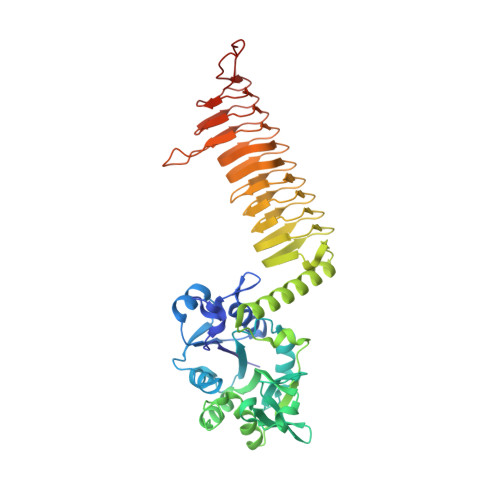
Structure of the E. coli bifunctional GlmU acetyltransferase active site with substrates and products.

Olsen, L.R.Vetting, M.W.Roderick, S.L.

(2007) Protein Sci 16: 1230-1235

  • DOI: https://doi.org/10.1110/ps.072779707
  • Primary Citation Related Structures: 
    2OI5, 2OI6, 2OI7

  • PubMed Abstract: 

    The biosynthesis of UDP-GlcNAc in bacteria is carried out by GlmU, an essential bifunctional uridyltransferase that catalyzes the CoA-dependent acetylation of GlcN-1-PO(4) to form GlcNAc-1-PO(4) and its subsequent condensation with UTP to form pyrophosphate and UDP-GlcNAc. As a metabolite, UDP-GlcNAc is situated at a branch point leading to the biosynthesis of lipopolysaccharide and peptidoglycan. Consequently, GlmU is regarded as an important target for potential antibacterial agents. The crystal structure of the Escherichia coli GlmU acetyltransferase active site has been determined in complexes with acetyl-CoA, CoA/GlcN-1-PO(4), and desulpho-CoA/GlcNAc-1-PO(4). These structures reveal the enzyme groups responsible for binding the substrates. A superposition of these complex structures suggests that the 2-amino group of GlcN-1-PO(4) is positioned in proximity to the acetyl-CoA to facilitate direct attack on its thioester by a ternary complex mechanism.


  • Organizational Affiliation
    • Department of Biochemistry, Albert Einstein College of Medicine, Bronx, New York 10461, USA.

Macromolecule Content 

  • Total Structure Weight: 101.75 kDa 
  • Atom Count: 7,590 
  • Modeled Residue Count: 899 
  • Deposited Residue Count: 912 
  • Unique protein chains: 1

Macromolecules

Find similar proteins by:|  3D Structure
Entity ID: 1
MoleculeChains  Sequence LengthOrganismDetailsImage
Bifunctional protein glmU
A, B
456Escherichia coliMutation(s): 0 
Gene Names: glmU
EC: 2.7.7.23 (PDB Primary Data), 2.3.1.157 (PDB Primary Data)
UniProt
Find proteins for P0ACC7 (Escherichia coli (strain K12))
Explore P0ACC7 
Go to UniProtKB:  P0ACC7
Entity Groups
Sequence Clusters30% Identity50% Identity70% Identity90% Identity95% Identity100% Identity
UniProt GroupP0ACC7
Sequence Annotations
Expand
Reference Sequence

Small Molecules

Ligands 4 Unique
IDChains Name / Formula / InChI Key2D Diagram3D Interactions
ACO

Query on ACO


Download:Ideal Coordinates CCD File
F [auth A],
M [auth B]
ACETYL COENZYME *A
C23 H38 N7 O17 P3 S
ZSLZBFCDCINBPY-ZSJPKINUSA-N
UD1

Query on UD1


Download:Ideal Coordinates CCD File
G [auth A],
N [auth B]
URIDINE-DIPHOSPHATE-N-ACETYLGLUCOSAMINE
C17 H27 N3 O17 P2
LFTYTUAZOPRMMI-CFRASDGPSA-N
SO4

Query on SO4


Download:Ideal Coordinates CCD File
E [auth A],
K [auth B],
L [auth B]
SULFATE ION
O4 S
QAOWNCQODCNURD-UHFFFAOYSA-L
MG

Query on MG


Download:Ideal Coordinates CCD File
C [auth A],
D [auth A],
H [auth B],
I [auth B],
J [auth B]
MAGNESIUM ION
Mg
JLVVSXFLKOJNIY-UHFFFAOYSA-N

Experimental Data & Validation

Experimental Data

  • Method: X-RAY DIFFRACTION
  • Resolution: 2.25 Å
  • R-Value Free:  0.219 (Depositor), 0.219 (DCC) 
  • R-Value Work:  0.176 (Depositor), 0.177 (DCC) 
  • R-Value Observed: 0.176 (Depositor) 
Space Group: H 3 2
Unit Cell:
Length ( Å )Angle ( ˚ )
a = 102.487α = 90
b = 102.487β = 90
c = 644.426γ = 120
Software Package:
Software NamePurpose
CNSrefinement
DENZOdata reduction
SCALEPACKdata scaling
CNSphasing

Structure Validation

View Full Validation Report


Ligand Structure Quality Assessment 


Entry History 

Deposition Data

Revision History  (Full details and data files)

  • Version 1.0: 2007-06-19
    Type: Initial release
  • Version 1.1: 2008-05-01
    Changes: Version format compliance
  • Version 1.2: 2011-07-13
    Changes: Version format compliance
  • Version 1.3: 2023-08-30
    Changes: Data collection, Database references, Derived calculations, Refinement description