RCSB PDB - 2RKZ: Crystal structure of the second and third fibronectin f1 modules in complex with a fragment of staphylococcus aureus fnbpa-1

 2RKZ

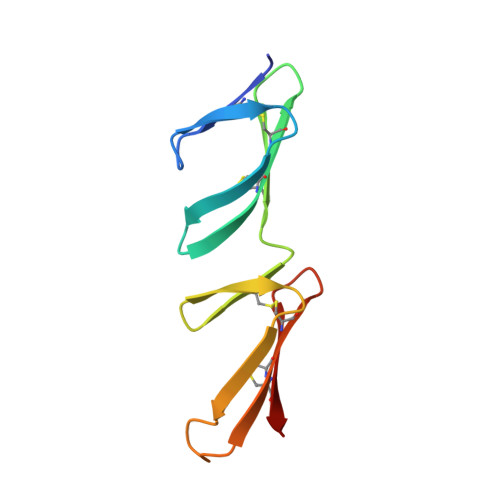
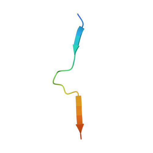
Crystal structure of the second and third fibronectin f1 modules in complex with a fragment of staphylococcus aureus fnbpa-1


Experimental Data Snapshot

  • Method: X-RAY DIFFRACTION
  • Resolution: 2.00 Å
  • R-Value Free: 0.261 
  • R-Value Work: 0.197 
  • R-Value Observed: 0.200 

Starting Model: experimental
View more details

wwPDB Validation   3D Report Full Report


This is version 2.2 of the entry. See complete history


Literature

Crystal structures of fibronectin-binding sites from Staphylococcus aureus FnBPA in complex with fibronectin domains

Bingham, R.J.Meenan, N.A.Schwarz-Linek, U.Turkenburg, J.P.Garman, E.F.Potts, J.R.

(2008) Proc Natl Acad Sci U S A 105: 12254-12258

  • DOI: https://doi.org/10.1073/pnas.0803556105
  • Primary Citation of Related Structures:  
    2RKY, 2RKZ, 2RL0, 3CAL

  • PubMed Abstract: 

    Staphylococcus aureus can adhere to and invade endothelial cells by binding to the human protein fibronectin (Fn). FnBPA and FnBPB, cell wall-attached proteins from S. aureus, have multiple, intrinsically disordered, high-affinity binding repeats (FnBRs) for Fn. Here, 30 years after the first report of S. aureus/Fn interactions, we present four crystal structures that together comprise the structures of two complete FnBRs, each in complex with four of the N-terminal modules of Fn. Each approximately 40-residue FnBR forms antiparallel strands along the triple-stranded beta-sheets of four sequential F1 modules ((2-5)F1) with each FnBR/(2-5)F1 interface burying a total surface area of approximately 4,300 A(2). The structures reveal the roles of residues conserved between S. aureus and Streptococcus pyogenes FnBRs and show that there are few linker residues between FnBRs. The ability to form large intermolecular interfaces with relatively few residues has been proposed to be a feature of disordered proteins, and S. aureus/Fn interactions provide an unusual illustration of this efficiency.


  • Organizational Affiliation

    Department of Biology, University of York, PO Box 373, York YO10 5YW, United Kingdom.


Macromolecules
Find similar proteins by:  (by identity cutoff)  |  3D Structure
Entity ID: 1
MoleculeChains Sequence LengthOrganismDetailsImage
Fibronectin
A, B, C, D, E
90Homo sapiensMutation(s): 0 
Gene Names: FN1
UniProt & NIH Common Fund Data Resources
Find proteins for P02751 (Homo sapiens)
Explore P02751 
Go to UniProtKB:  P02751
PHAROS:  P02751
GTEx:  ENSG00000115414 
Entity Groups  
Sequence Clusters30% Identity50% Identity70% Identity90% Identity95% Identity100% Identity
UniProt GroupP02751
Sequence Annotations
Expand
  • Reference Sequence

Find similar proteins by:  Sequence   |   3D Structure  

Entity ID: 2
MoleculeChains Sequence LengthOrganismDetailsImage
peptide from Fibronectin-binding protein A23Staphylococcus aureus subsp. aureus NCTC 8325Mutation(s): 2 
UniProt
Find proteins for P14738 (Staphylococcus aureus (strain NCTC 8325 / PS 47))
Explore P14738 
Go to UniProtKB:  P14738
Entity Groups  
Sequence Clusters30% Identity50% Identity70% Identity90% Identity95% Identity100% Identity
UniProt GroupP14738
Sequence Annotations
Expand
  • Reference Sequence
Experimental Data & Validation

Experimental Data

  • Method: X-RAY DIFFRACTION
  • Resolution: 2.00 Å
  • R-Value Free: 0.261 
  • R-Value Work: 0.197 
  • R-Value Observed: 0.200 
  • Space Group: P 1 21 1
Unit Cell:
Length ( Å )Angle ( ˚ )
a = 59.59α = 90
b = 109.72β = 96.94
c = 71.47γ = 90
Software Package:
Software NamePurpose
SCALAdata scaling
REFMACrefinement
PDB_EXTRACTdata extraction
ADSCdata collection
MOSFLMdata reduction
PHASERphasing

Structure Validation

View Full Validation Report


Entry History 

Deposition Data

Revision History  (Full details and data files)

  • Version 1.0: 2008-08-05
    Type: Initial release
  • Version 1.1: 2011-07-13
    Changes: Version format compliance
  • Version 2.0: 2019-06-19
    Changes: Advisory, Atomic model, Data collection, Database references, Derived calculations, Polymer sequence, Source and taxonomy, Structure summary
  • Version 2.1: 2024-04-03
    Changes: Data collection, Database references, Derived calculations, Refinement description
  • Version 2.2: 2024-11-13
    Changes: Structure summary