2WH5

Crystal structure of human acyl-CoA binding domain 4 complexed with stearoyl-CoA


Experimental Data Snapshot

  • Method: X-RAY DIFFRACTION
  • Resolution: 2.60 Å
  • R-Value Free: 
    0.236 (Depositor), 0.220 (DCC) 
  • R-Value Work: 
    0.206 (Depositor), 0.190 (DCC) 
  • R-Value Observed: 
    0.207 (Depositor) 

Starting Model: experimental
View more details

wwPDB Validation   3D Report Full Report


Ligand Structure Quality Assessment 

Created with Raphaël 2.3.0Worse 01 BetterLigand structure goodness of fit to experimental dataBest fitted ST9Click on this verticalbar to view detailsBest fitted COAClick on this verticalbar to view detailsBest fitted STEClick on this verticalbar to view details

This is version 1.3 of the entry. See complete history


Literature

Crystal Structure of Human Acyl-Coa Binding Domain 4

Shafqat, N.Yue, W.W.Oppermann, U.

To be published.

Macromolecules
Find similar proteins by:  (by identity cutoff)  |  3D Structure
Entity ID: 1
MoleculeChains Sequence LengthOrganismDetailsImage
ACYL-COA-BINDING DOMAIN-CONTAINING PROTEIN 4
A, B, C, D, E
106Homo sapiensMutation(s): 0 
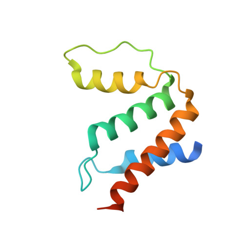
UniProt & NIH Common Fund Data Resources
Find proteins for Q8NC06 (Homo sapiens)
Explore Q8NC06 
Go to UniProtKB:  Q8NC06
PHAROS:  Q8NC06
GTEx:  ENSG00000181513 
Entity Groups  
Sequence Clusters30% Identity50% Identity70% Identity90% Identity95% Identity100% Identity
UniProt GroupQ8NC06
Sequence Annotations
Expand
  • Reference Sequence
Small Molecules
Ligands 3 Unique
IDChains Name / Formula / InChI Key2D Diagram3D Interactions
ST9
Query on ST9

Download Ideal Coordinates CCD File 
H [auth A]
J [auth B]
L [auth C]
N [auth D]
Q [auth E]
STEAROYL-COENZYME A
C39 H70 N7 O17 P3 S
SIARJEKBADXQJG-LFZQUHGESA-N
COA
Query on COA

Download Ideal Coordinates CCD File 
O [auth D],
R [auth E]
COENZYME A
C21 H36 N7 O16 P3 S
RGJOEKWQDUBAIZ-IBOSZNHHSA-N
STE
Query on STE

Download Ideal Coordinates CCD File 
G [auth A]
I [auth B]
K [auth C]
M [auth D]
P [auth E]
STEARIC ACID
C18 H36 O2
QIQXTHQIDYTFRH-UHFFFAOYSA-N
Experimental Data & Validation

Experimental Data

  • Method: X-RAY DIFFRACTION
  • Resolution: 2.60 Å
  • R-Value Free:  0.236 (Depositor), 0.220 (DCC) 
  • R-Value Work:  0.206 (Depositor), 0.190 (DCC) 
  • R-Value Observed: 0.207 (Depositor) 
Space Group: P 1 21 1
Unit Cell:
Length ( Å )Angle ( ˚ )
a = 85.06α = 90
b = 95.1β = 110.28
c = 119.74γ = 90
Software Package:
Software NamePurpose
PHENIXrefinement
MOSFLMdata reduction
SCALAdata scaling
PHASERphasing

Structure Validation

View Full Validation Report


Ligand Structure Quality Assessment 

Created with Raphaël 2.3.0Worse 01 BetterLigand structure goodness of fit to experimental dataBest fitted ST9Click on this verticalbar to view detailsBest fitted COAClick on this verticalbar to view detailsBest fitted STEClick on this verticalbar to view details

Entry History 

Revision History  (Full details and data files)

  • Version 1.0: 2009-07-28
    Type: Initial release
  • Version 1.1: 2012-12-05
    Changes: Database references, Derived calculations, Other, Refinement description, Structure summary, Version format compliance
  • Version 1.2: 2013-09-25
    Changes: Advisory, Non-polymer description, Source and taxonomy
  • Version 1.3: 2023-12-13
    Changes: Data collection, Database references, Derived calculations, Other, Refinement description