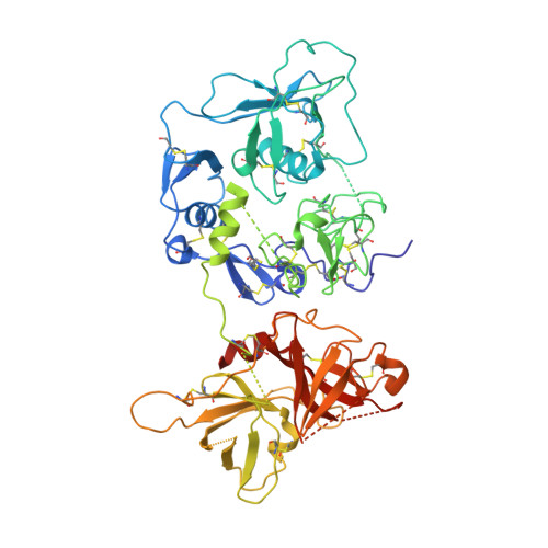
2XRC

Human complement factor I


Experimental Data Snapshot

  • Method: X-RAY DIFFRACTION
  • Resolution: 2.69 Å
  • R-Value Free: 0.238 
  • R-Value Work: 0.200 
  • R-Value Observed: 0.202 

Starting Models: experimental
View more details

wwPDB Validation   3D Report Full Report


Ligand Structure Quality Assessment 


This is version 1.3 of the entry. See complete history


Literature

Structural Basis for Complement Factor I Control and its Disease-Associated Sequence Polymorphisms.

Roversi, P.Johnson, S.Caesar, J.J.Mclean, F.Leath, K.J.Tsiftsoglou, S.A.Morgan, B.P.Harris, C.L.Sim, R.B.Lea, S.M.

(2011) Proc Natl Acad Sci U S A 108: 12839

  • DOI: https://doi.org/10.1073/pnas.1102167108
  • Primary Citation of Related Structures:  
    2XRC

  • PubMed Abstract: 

    The complement system is a key component of innate and adaptive immune responses. Complement regulation is critical for prevention and control of disease. We have determined the crystal structure of the complement regulatory enzyme human factor I (fI). FI is in a proteolytically inactive form, demonstrating that it circulates in a zymogen-like state despite being fully processed to the mature sequence. Mapping of functional data from mutants of fI onto the structure suggests that this inactive form is maintained by the noncatalytic heavy-chain allosterically modulating activity of the light chain. Once the ternary complex of fI, a cofactor and a substrate is formed, the allosteric inhibition is released, and fI is oriented for cleavage. In addition to explaining how circulating fI is limited to cleaving only C3b/C4b, our model explains the molecular basis of disease-associated polymorphisms in fI and its cofactors.


  • Organizational Affiliation

    Sir William Dunn School of Pathology, University of Oxford, Oxford OX1 3RE, United Kingdom.


Macromolecules
Find similar proteins by:  (by identity cutoff)  |  3D Structure
Entity ID: 1
MoleculeChains Sequence LengthOrganismDetailsImage
HUMAN COMPLEMENT FACTOR I
A, B, C, D
565Homo sapiensMutation(s): 0 
EC: 3.4.21.45
UniProt & NIH Common Fund Data Resources
Find proteins for P05156 (Homo sapiens)
Explore P05156 
Go to UniProtKB:  P05156
PHAROS:  P05156
GTEx:  ENSG00000205403 
Entity Groups  
Sequence Clusters30% Identity50% Identity70% Identity90% Identity95% Identity100% Identity
UniProt GroupP05156
Glycosylation
Glycosylation Sites: 6Go to GlyGen: P05156-1
Sequence Annotations
Expand
  • Reference Sequence
Small Molecules
Ligands 2 Unique
IDChains Name / Formula / InChI Key2D Diagram3D Interactions
NAG
Query on NAG

Download Ideal Coordinates CCD File 
AA [auth C]
DA [auth D]
EA [auth D]
FA [auth D]
G [auth A]
AA [auth C],
DA [auth D],
EA [auth D],
FA [auth D],
G [auth A],
GA [auth D],
H [auth A],
HA [auth D],
I [auth A],
J [auth A],
K [auth A],
L [auth A],
O [auth B],
P [auth B],
Q [auth B],
R [auth B],
S [auth B],
T [auth B],
W [auth C],
X [auth C],
Y [auth C],
Z [auth C]
2-acetamido-2-deoxy-beta-D-glucopyranose
C8 H15 N O6
OVRNDRQMDRJTHS-FMDGEEDCSA-N
CA
Query on CA

Download Ideal Coordinates CCD File 
BA [auth D]
CA [auth D]
E [auth A]
F [auth A]
M [auth B]
BA [auth D],
CA [auth D],
E [auth A],
F [auth A],
M [auth B],
N [auth B],
U [auth C],
V [auth C]
CALCIUM ION
Ca
BHPQYMZQTOCNFJ-UHFFFAOYSA-N
Experimental Data & Validation

Experimental Data

  • Method: X-RAY DIFFRACTION
  • Resolution: 2.69 Å
  • R-Value Free: 0.238 
  • R-Value Work: 0.200 
  • R-Value Observed: 0.202 
  • Space Group: P 1
Unit Cell:
Length ( Å )Angle ( ˚ )
a = 71.317α = 89.98
b = 234.721β = 90.18
c = 40.3γ = 90.03
Software Package:
Software NamePurpose
REFMACrefinement
XDSdata reduction
SCALAdata scaling
PHASERphasing

Structure Validation

View Full Validation Report


Ligand Structure Quality Assessment 


Entry History 

Deposition Data

Revision History  (Full details and data files)

  • Version 1.0: 2011-07-13
    Type: Initial release
  • Version 1.1: 2011-08-17
    Changes: Database references
  • Version 1.2: 2020-07-29
    Type: Remediation
    Reason: Carbohydrate remediation
    Changes: Advisory, Data collection, Derived calculations, Other, Structure summary
  • Version 1.3: 2023-12-20
    Changes: Data collection, Database references, Derived calculations, Refinement description, Structure summary