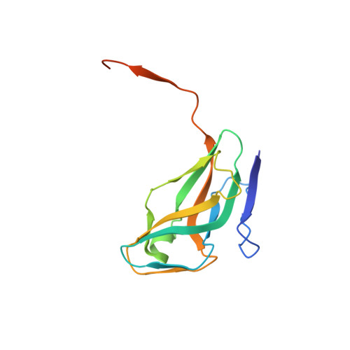
The Structure of Bacillus Subtilis Sp Beta Prophage Dutpase and its Complexes with Two Nucleotides
Garcia-Nafria, J., Harkiolaki, M., Persson, R., Fogg, M.J., Wilson, K.S.(2011) Acta Crystallogr D Biol Crystallogr 67: 167
- PubMed: 21358047
- DOI: https://doi.org/10.1107/S0907444911003234
- Primary Citation of Related Structures:
2XX6, 2XY3, 2Y1T - PubMed Abstract:
dUTPases are housekeeping enzymes which catalyse the hydrolysis of dUTP to dUMP in an ion-dependent manner. Bacillus subtilis has both a genomic and an SPβ prophage homotrimeric dUTPase. Here, structure determination of the prophage apoenzyme and of its complexes with dUDP and dUpNHpp-Mg(2+) is described at 1.75, 1.9 and 2.55 Å resolution, respectively. The C-terminal extension, which carries the conserved motif V, is disordered in all three structures. Unlike all other trimeric dUTPases for which structures are available, with the exception of the Bacillus genomic enzyme, the aromatic residue covering the uridine and acting as the Phe-lid is close to motif III in the sequence rather than in motif V. This is in spite of the presence of an aromatic amino acid at the usual Phe-lid position in motif V. The alternative position of the Phe-lid requires a reconsideration of its role in the catalytic cycle of the enzyme. In the dUpNHpp-Mg(2+) complex a water can be seen at the position expected for nucleophilic attack on the α-phosphate, in spite of motif V being disordered. Differences in the active site between the free enzyme and the dUDP and dUpNHpp-Mg(2+) complexes shows that the triphosphate moiety needs to be in the gauche conformation to trigger the conformational changes that can be seen in both B. subtilis dUTPases.
Organizational Affiliation:
Department of Chemistry, University of York, York, England.


