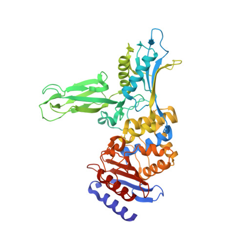
2Y55

Unexpected tricovalent binding mode of boronic acids within the active site of a penicillin binding protein


Experimental Data Snapshot

  • Method: X-RAY DIFFRACTION
  • Resolution: 2.60 Å
  • R-Value Free: 0.262 
  • R-Value Work: 0.194 
  • R-Value Observed: 0.197 

Starting Model: experimental
View more details

wwPDB Validation   3D Report Full Report


Ligand Structure Quality Assessment 


This is version 1.1 of the entry. See complete history


Literature

Unexpected Tricovalent Binding Mode of Boronic Acids within the Active Site of a Penicillin- Binding Protein.

Zervosen, A.Herman, R.Kerff, F.Herman, A.Bouillez, A.Prati, F.Pratt, R.F.Frere, J.M.Joris, B.Luxen, A.Charlier, P.Sauvage, E.

(2011) J Am Chem Soc 133: 10839

  • DOI: https://doi.org/10.1021/ja200696y
  • Primary Citation of Related Structures:  
    2Y4A, 2Y55, 2Y59, 3ZVT, 3ZVW

  • PubMed Abstract: 

    Boronic acids bearing appropriate side chains are good inhibitors of serine amidohydrolases. The boron usually adopts a tetrahedral conformation, bound to the nucleophilic serine of the active site and mimicking the transition state of the enzymatic reaction. We have solved the structures of complexes of a penicillin-binding protein, the DD-peptidase from Actinomadura sp. R39, with four amidomethylboronic acids (2,6-dimethoxybenzamidomethylboronic acid, phenylacetamidomethylboronic acid, 2-chlorobenzamidomethylboronic acid, and 2-nitrobenzamidomethylboronic acid) and the pinacol ester derived from phenylacetamidomethylboronic acid. We found that, in each case, the boron forms a tricovalent adduct with Oγ of Ser49, Ser298, and the terminal amine group of Lys410, three key residues involved in the catalytic mechanism of penicillin-binding proteins. This represents the first tricovalent enzyme-inhibitor adducts observed by crystallography. In two of the five R39-boronate structures, the boronic acid is found as a tricovalent adduct in two monomers of the asymmetric unit and as a monocovalent adduct with the active serine in the two remaining monomers of the asymmetric unit. Formation of the tricovalent complex from a classical monocovalent complex may involve rotation around the Ser49 Cα-Cβ bond to place the boron in a position to interact with Ser298 and Lys410, and a twisting of the side-chain amide such that its carbonyl oxygen is able to hydrogen bond to the oxyanion hole NH of Thr413. Biphasic kinetics were observed in three of the five cases, and details of the reaction between R39 and 2,6-dimethoxybenzamidomethylboronic acid were studied. Observation of biphasic kinetics was not, however, thought to be correlated to formation of tricovalent complexes, assuming that the latter do form in solution. On the basis of the crystallographic and kinetic results, a reaction scheme for this unexpected inhibition by boronic acids is proposed.


  • Organizational Affiliation

    Centre de Recherches du Cyclotron, Université de Liège, B-4000 Sart Tilman, Liège, Belgium.


Macromolecules
Find similar proteins by:  (by identity cutoff)  |  3D Structure
Entity ID: 1
MoleculeChains Sequence LengthOrganismDetailsImage
D-ALANYL-D-ALANINE CARBOXYPEPTIDASE
A, B, C, D
466Actinomadura sp. R39Mutation(s): 0 
EC: 3.4.16.4
UniProt
Find proteins for P39045 (Actinomadura sp. (strain R39))
Explore P39045 
Go to UniProtKB:  P39045
Entity Groups  
Sequence Clusters30% Identity50% Identity70% Identity90% Identity95% Identity100% Identity
UniProt GroupP39045
Sequence Annotations
Expand
  • Reference Sequence
Small Molecules
Ligands 4 Unique
IDChains Name / Formula / InChI Key2D Diagram3D Interactions
FP5
Query on FP5

Download Ideal Coordinates CCD File 
E [auth A],
N [auth B],
T [auth C],
Z [auth D]
PHENYLACETAMIDOMETHYL BORONIC ACID
C9 H12 B N O3
WPZJZIUTEZTIJI-UHFFFAOYSA-N
SO4
Query on SO4

Download Ideal Coordinates CCD File 
AA [auth D]
BA [auth D]
CA [auth D]
DA [auth D]
F [auth A]
AA [auth D],
BA [auth D],
CA [auth D],
DA [auth D],
F [auth A],
G [auth A],
H [auth A],
I [auth A],
M [auth A],
O [auth B],
P [auth B],
Q [auth B],
R [auth B],
U [auth C],
V [auth C],
W [auth C],
X [auth C]
SULFATE ION
O4 S
QAOWNCQODCNURD-UHFFFAOYSA-L
ACN
Query on ACN

Download Ideal Coordinates CCD File 
GA [auth D],
HA [auth D],
L [auth A],
S [auth B],
Y [auth C]
ACETONE
C3 H6 O
CSCPPACGZOOCGX-UHFFFAOYSA-N
MG
Query on MG

Download Ideal Coordinates CCD File 
EA [auth D],
FA [auth D],
J [auth A],
K [auth A]
MAGNESIUM ION
Mg
JLVVSXFLKOJNIY-UHFFFAOYSA-N
Binding Affinity Annotations 
IDSourceBinding Affinity
FP5 BindingDB:  2Y55 Ki: 1.25e+5 (nM) from 1 assay(s)
IC50: 3.20e+5 (nM) from 1 assay(s)
PDBBind:  2Y55 Ki: 1.34e+5 (nM) from 1 assay(s)
Experimental Data & Validation

Experimental Data

  • Method: X-RAY DIFFRACTION
  • Resolution: 2.60 Å
  • R-Value Free: 0.262 
  • R-Value Work: 0.194 
  • R-Value Observed: 0.197 
  • Space Group: P 1 21 1
Unit Cell:
Length ( Å )Angle ( ˚ )
a = 104.234α = 90
b = 92.284β = 94.2
c = 107.105γ = 90
Software Package:
Software NamePurpose
REFMACrefinement
MOSFLMdata reduction
SCALAdata scaling

Structure Validation

View Full Validation Report


Ligand Structure Quality Assessment 


Entry History 

Deposition Data

Revision History  (Full details and data files)

  • Version 1.0: 2011-07-27
    Type: Initial release
  • Version 1.1: 2023-12-20
    Changes: Data collection, Database references, Derived calculations, Other, Refinement description