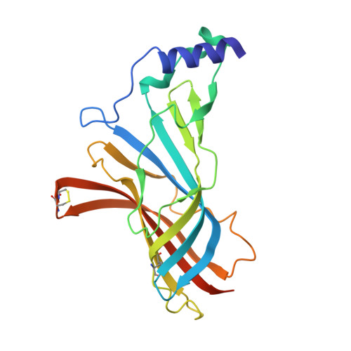
2Y56

Fragment growing induces conformational changes in acetylcholine- binding protein: A structural and thermodynamic analysis - (Compound 3)


Experimental Data Snapshot

  • Method: X-RAY DIFFRACTION
  • Resolution: 3.59 Å
  • R-Value Free: 0.203 
  • R-Value Work: 0.174 
  • R-Value Observed: 0.175 

Starting Model: experimental
View more details

wwPDB Validation   3D Report Full Report


Ligand Structure Quality Assessment 


This is version 1.4 of the entry. See complete history


Literature

Fragment Growing Induces Conformational Changes in Acetylcholine-Binding Protein: A Structural and Thermodynamic Analysis.

Edink, E.Rucktooa, P.Retra, K.Akdemir, A.Nahar, T.Zuiderveld, O.Van Elk, R.Janssen, E.Van Nierop, P.Van Muijlwijk-Koezen, J.Smit, A.B.Sixma, T.K.Leurs, R.De Esch, I.J.P.

(2011) J Am Chem Soc 133: 5363

  • DOI: https://doi.org/10.1021/ja110571r
  • Primary Citation of Related Structures:  
    2Y54, 2Y56, 2Y57, 2Y58

  • PubMed Abstract: 

    Optimization of fragment hits toward high-affinity lead compounds is a crucial aspect of fragment-based drug discovery (FBDD). In the current study, we have successfully optimized a fragment by growing into a ligand-inducible subpocket of the binding site of acetylcholine-binding protein (AChBP). This protein is a soluble homologue of the ligand binding domain (LBD) of Cys-loop receptors. The fragment optimization was monitored with X-ray structures of ligand complexes and systematic thermodynamic analyses using surface plasmon resonance (SPR) biosensor analysis and isothermal titration calorimetry (ITC). Using site-directed mutagenesis and AChBP from different species, we find that specific changes in thermodynamic binding profiles, are indicative of interactions with the ligand-inducible subpocket of AChBP. This study illustrates that thermodynamic analysis provides valuable information on ligand binding modes and is complementary to affinity data when guiding rational structure- and fragment-based discovery approaches.


  • Organizational Affiliation

    Leiden/Amsterdam Center of Drug Research (LACDR), Division of Medicinal Chemistry, Faculty of Sciences, VU University Amsterdam, The Netherlands.


Macromolecules
Find similar proteins by:  (by identity cutoff)  |  3D Structure
Entity ID: 1
MoleculeChains Sequence LengthOrganismDetailsImage
SOLUBLE ACETYLCHOLINE RECEPTOR
A, B, C, D, E
217Aplysia californicaMutation(s): 0 
UniProt
Find proteins for Q8WSF8 (Aplysia californica)
Explore Q8WSF8 
Go to UniProtKB:  Q8WSF8
Entity Groups  
Sequence Clusters30% Identity50% Identity70% Identity90% Identity95% Identity100% Identity
UniProt GroupQ8WSF8
Sequence Annotations
Expand
  • Reference Sequence
Small Molecules
Ligands 4 Unique
IDChains Name / Formula / InChI Key2D Diagram3D Interactions
V11
Query on V11

Download Ideal Coordinates CCD File 
AA [auth D],
F [auth A],
KA [auth E],
M [auth B],
T [auth C]
[(1S,5R)-8-[(2R)-2-HYDROXY-2-PHENYL-ETHYL]-8-AZABICYCLO[3.2.1]OCTAN-3-YL] BENZOATE
C22 H25 N O3
CAELFFAMXVUOKR-BQJUDKOJSA-N
SO4
Query on SO4

Download Ideal Coordinates CCD File 
EA [auth D]
FA [auth D]
GA [auth D]
I [auth A]
J [auth A]
EA [auth D],
FA [auth D],
GA [auth D],
I [auth A],
J [auth A],
NA [auth E],
OA [auth E],
PA [auth E],
Q [auth B],
QA [auth E],
R [auth B],
RA [auth E],
W [auth C],
X [auth C]
SULFATE ION
O4 S
QAOWNCQODCNURD-UHFFFAOYSA-L
GOL
Query on GOL

Download Ideal Coordinates CCD File 
HA [auth D]
IA [auth D]
JA [auth D]
K [auth A]
L [auth A]
HA [auth D],
IA [auth D],
JA [auth D],
K [auth A],
L [auth A],
S [auth B],
Y [auth C],
Z [auth C]
GLYCEROL
C3 H8 O3
PEDCQBHIVMGVHV-UHFFFAOYSA-N
CL
Query on CL

Download Ideal Coordinates CCD File 
BA [auth D]
CA [auth D]
DA [auth D]
G [auth A]
H [auth A]
BA [auth D],
CA [auth D],
DA [auth D],
G [auth A],
H [auth A],
LA [auth E],
MA [auth E],
N [auth B],
O [auth B],
P [auth B],
U [auth C],
V [auth C]
CHLORIDE ION
Cl
VEXZGXHMUGYJMC-UHFFFAOYSA-M
Binding Affinity Annotations 
IDSourceBinding Affinity
V11 BindingDB:  2Y56 Ki: 100 (nM) from 1 assay(s)
PDBBind:  2Y56 Kd: 310 (nM) from 1 assay(s)
Experimental Data & Validation

Experimental Data

  • Method: X-RAY DIFFRACTION
  • Resolution: 3.59 Å
  • R-Value Free: 0.203 
  • R-Value Work: 0.174 
  • R-Value Observed: 0.175 
  • Space Group: I 2 3
Unit Cell:
Length ( Å )Angle ( ˚ )
a = 218.93α = 90
b = 218.93β = 90
c = 218.93γ = 90
Software Package:
Software NamePurpose
BUSTERrefinement
XDSdata reduction
XSCALEdata scaling
PHASERphasing

Structure Validation

View Full Validation Report


Ligand Structure Quality Assessment 


Entry History 

Deposition Data

Revision History  (Full details and data files)

  • Version 1.0: 2011-06-15
    Type: Initial release
  • Version 1.1: 2011-07-13
    Changes: Version format compliance
  • Version 1.2: 2019-04-24
    Changes: Data collection, Other, Source and taxonomy
  • Version 1.3: 2023-12-20
    Changes: Data collection, Database references, Derived calculations, Other, Refinement description
  • Version 1.4: 2024-10-16
    Changes: Structure summary