2Z5O | pdb_00002z5o

Complex of Transportin 1 with JKTBP NLS


Experimental Data Snapshot

  • Method: X-RAY DIFFRACTION
  • Resolution: 3.20 Å
  • R-Value Free: 
    0.279 (Depositor), 0.224 (DCC) 
  • R-Value Work: 
    0.236 (Depositor), 0.229 (DCC) 
  • R-Value Observed: 
    0.238 (Depositor) 

Starting Model: experimental
View more details

wwPDB Validation 3D Report Full Report

Validation slider image for 2Z5O

This is version 1.2 of the entry. See complete history

Literature

Structural basis for substrate recognition and dissociation by human transportin 1

Imasaki, T.Shimizu, T.Hashimoto, H.Hidaka, Y.Kose, S.Imamoto, N.Yamada, M.Sato, M.

(2007) Mol Cell 28: 57-67

  • DOI: https://doi.org/10.1016/j.molcel.2007.08.006
  • Primary Citation Related Structures: 
    2Z5J, 2Z5K, 2Z5M, 2Z5N, 2Z5O

  • PubMed Abstract: 

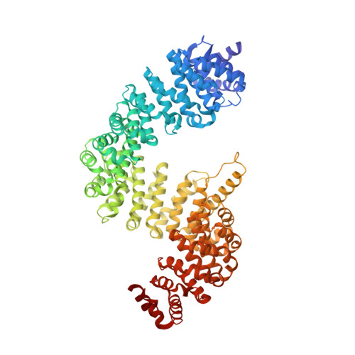
    Transportin 1 (Trn1) is a transport receptor that transports substrates from the cytoplasm to the nucleus through nuclear pore complexes by recognizing nuclear localization signals (NLSs). Here we describe four crystal structures of human Trn1 in a substrate-free form as well as in the complex with three NLSs (hnRNP D, JKTBP, and TAP, respectively). Our data have revealed that (1) Trn1 has two sites for binding NLSs, one with high affinity (site A) and one with low affinity (site B), and NLS interaction at site B controls overall binding affinity for Trn1; (2) Trn1 recognizes the NLSs at site A followed by conformational change at site B to interact with the NLSs; and (3) a long flexible loop, characteristic of Trn1, interacts with site B, thereby displacing transport substrate in the nucleus. These studies provide deep understanding of substrate recognition and dissociation by Trn1 in import pathways.


  • Organizational Affiliation
    • Field of Supramolecular Biology, International Graduate School of Arts and Sciences, Yokohama City University, 1-7-29 Suehiro-cho, Tsurumi-ku, Yokohama 230-0045, Japan.

Macromolecule Content 

  • Total Structure Weight: 102.28 kDa 
  • Atom Count: 6,689 
  • Modeled Residue Count: 844 
  • Deposited Residue Count: 900 
  • Unique protein chains: 2

Macromolecules

Find similar proteins by:|  3D Structure
Entity ID: 1
MoleculeChains  Sequence LengthOrganismDetailsImage
Transportin-1890Homo sapiensMutation(s): 0 
UniProt & NIH Common Fund Data Resources
Find proteins for Q92973 (Homo sapiens)
Explore Q92973 
Go to UniProtKB:  Q92973
PHAROS:  Q92973
GTEx:  ENSG00000083312 
Entity Groups
Sequence Clusters30% Identity50% Identity70% Identity90% Identity95% Identity100% Identity
UniProt GroupQ92973
Sequence Annotations
Expand
Reference Sequence
Find similar proteins by:  Sequence   |   3D Structure  
Entity ID: 2
MoleculeChains  Sequence LengthOrganismDetailsImage
Heterogeneous nuclear ribonucleoprotein D-like10N/AMutation(s): 0 

Experimental Data & Validation

Experimental Data

  • Method: X-RAY DIFFRACTION
  • Resolution: 3.20 Å
  • R-Value Free:  0.279 (Depositor), 0.224 (DCC) 
  • R-Value Work:  0.236 (Depositor), 0.229 (DCC) 
  • R-Value Observed: 0.238 (Depositor) 
Space Group: P 21 21 2
Unit Cell:
Length ( Å )Angle ( ˚ )
a = 132.306α = 90
b = 169.805β = 90
c = 68.458γ = 90
Software Package:
Software NamePurpose
REFMACrefinement
ADSCdata collection
HKL-2000data reduction
HKL-2000data scaling
PHASERphasing

Structure Validation

View Full Validation Report


Entry History 

Deposition Data

Revision History  (Full details and data files)

  • Version 1.0: 2007-10-23
    Type: Initial release
  • Version 1.1: 2011-07-13
    Changes: Advisory, Version format compliance
  • Version 1.2: 2023-11-01
    Changes: Data collection, Database references, Refinement description