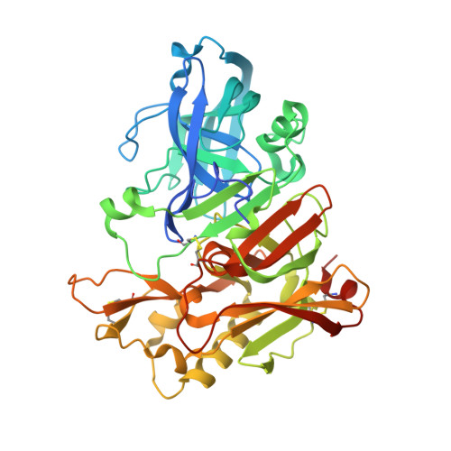
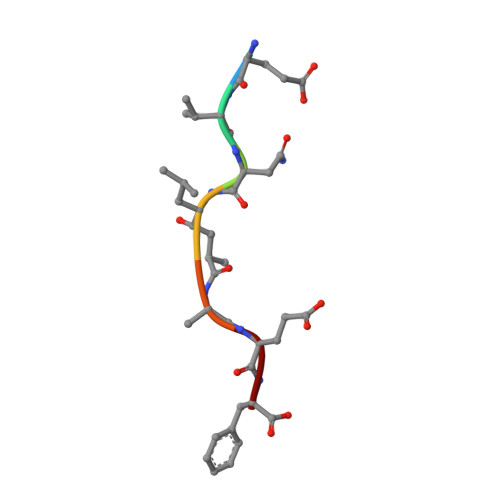
2ZHR

Crystal structure of BACE1 in complex with OM99-2 at pH 5.0


Experimental Data Snapshot

  • Method: X-RAY DIFFRACTION
  • Resolution: 2.50 Å
  • R-Value Free: 0.240 
  • R-Value Work: 0.185 
  • R-Value Observed: 0.185 

Starting Model: experimental
View more details

wwPDB Validation   3D Report Full Report


This is version 2.0 of the entry. See complete history


Literature

Crystal structure of an active form of BACE1, an enzyme responsible for amyloid beta protein production

Shimizu, H.Tosaki, A.Kaneko, K.Hisano, T.Sakurai, T.Nukina, N.

(2008) Mol Cell Biol 28: 3663-3671

  • DOI: https://doi.org/10.1128/MCB.02185-07
  • Primary Citation of Related Structures:  
    2ZHR, 2ZHS, 2ZHT, 2ZHU, 2ZHV

  • PubMed Abstract: 

    BACE1 (beta-secretase) is a transmembrane aspartic protease that cleaves the beta-amyloid precursor protein and generates the amyloid beta peptide (Abeta). BACE1 cycles between the cell surface and the endosomal system many times and becomes activated interconvertibly during its cellular trafficking, leading to the production of Abeta. Here we report the crystal structure of the catalytically active form of BACE1. The active form has novel structural features involving the conformation of the flap and subsites that promote substrate binding. The functionally essential residues and water molecules are well defined and play a key role in the iterative activation of BACE1. We further describe the crystal structure of the dehydrated form of BACE1, showing that BACE1 activity is dependent on the dynamics of a catalytically required Asp-bound water molecule, which directly affects its catalytic properties. These findings provide insight into a novel regulation of BACE1 activity and elucidate how BACE1 modulates its activity during cellular trafficking.


  • Organizational Affiliation

    Laboratory for Structural Neuropathology, RIKEN Brain Science Institute, 2-1 Hirosawa, Wako-shi, Saitama 351-0198, Japan.


Macromolecules
Find similar proteins by:  (by identity cutoff)  |  3D Structure
Entity ID: 1
MoleculeChains Sequence LengthOrganismDetailsImage
Beta-secretase 1
A, B
411Homo sapiensMutation(s): 0 
Gene Names: BACE1
EC: 3.4.23.46
UniProt & NIH Common Fund Data Resources
Find proteins for P56817 (Homo sapiens)
Explore P56817 
Go to UniProtKB:  P56817
PHAROS:  P56817
GTEx:  ENSG00000186318 
Entity Groups  
Sequence Clusters30% Identity50% Identity70% Identity90% Identity95% Identity100% Identity
UniProt GroupP56817
Sequence Annotations
Expand
  • Reference Sequence

Find similar proteins by:  Sequence   |   3D Structure  

Entity ID: 2
MoleculeChains Sequence LengthOrganismDetailsImage
inhibitor OM99-2
C, D
7N/AMutation(s): 0 
Sequence Annotations
Expand
  • Reference Sequence
Biologically Interesting Molecules (External Reference) 1 Unique
Experimental Data & Validation

Experimental Data

  • Method: X-RAY DIFFRACTION
  • Resolution: 2.50 Å
  • R-Value Free: 0.240 
  • R-Value Work: 0.185 
  • R-Value Observed: 0.185 
  • Space Group: P 21 21 21
Unit Cell:
Length ( Å )Angle ( ˚ )
a = 86.333α = 90
b = 89.912β = 90
c = 130.794γ = 90
Software Package:
Software NamePurpose
CNSrefinement
HKL-2000data reduction
SCALEPACKdata scaling
MOLREPphasing

Structure Validation

View Full Validation Report


Entry History 

Deposition Data

Revision History  (Full details and data files)

  • Version 1.0: 2008-04-22
    Type: Initial release
  • Version 1.1: 2011-07-13
    Changes: Atomic model, Database references, Derived calculations, Non-polymer description, Structure summary, Version format compliance
  • Version 1.2: 2012-12-12
    Changes: Other
  • Version 1.3: 2023-11-01
    Changes: Data collection, Database references, Derived calculations, Refinement description
  • Version 2.0: 2023-11-15
    Changes: Atomic model, Data collection