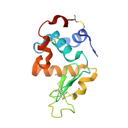
3B0I

Crystal structure of recombinant human alpha lactalbumin


Experimental Data Snapshot

  • Method: X-RAY DIFFRACTION
  • Resolution: 1.80 Å
  • R-Value Free: 0.256 
  • R-Value Work: 0.209 
  • R-Value Observed: 0.212 

Starting Model: experimental
View more details

wwPDB Validation   3D Report Full Report


This is version 1.2 of the entry. See complete history


Literature

Structural insights into the stability perturbations induced by N-terminal variation in human and goat alpha-lactalbumin

Makabe, K.Nakamura, T.Kuwajima, K.

(2013) Protein Eng Des Sel 26: 165-170

  • DOI: https://doi.org/10.1093/protein/gzs093
  • Primary Citation of Related Structures:  
    3B0I, 3B0K, 3B0O

  • PubMed Abstract: 

    Addition of an extra methionine at the N-terminus by recombinant expression of α-lactalbumin in Escherichia coli significantly destabilizes the protein, and this destabilization has hampered mutational analyses such as the mutational phi-value analysis of the protein. Deletion of residue 1 from the recombinant form recovers the stability in human and goat α-lactalbumin. Here, we thus determined the crystal structures of the residue 1-deletion variants of recombinant human and goat α-lactalbumin, and compared the structures with those of the authentic and recombinant forms. The results demonstrate the importance of the N-terminal backbone structure and hydrogen-bonding pattern for the stability of α-lactalbumin.


  • Organizational Affiliation

    Okazaki Institute for Integrative Bioscience and Institute for Molecular Science, National Institutes of Natural Sciences, 5-1 Higashiyama, Myodaiji, Okazaki 444-8787, Japan. makabe@yz.yamagata-u.ac.jp


Macromolecules
Find similar proteins by:  (by identity cutoff)  |  3D Structure
Entity ID: 1
MoleculeChains Sequence LengthOrganismDetailsImage
Alpha-lactalbumin124Homo sapiensMutation(s): 0 
Gene Names: LALBALYZL7
UniProt & NIH Common Fund Data Resources
Find proteins for P00709 (Homo sapiens)
Explore P00709 
Go to UniProtKB:  P00709
PHAROS:  P00709
GTEx:  ENSG00000167531 
Entity Groups  
Sequence Clusters30% Identity50% Identity70% Identity90% Identity95% Identity100% Identity
UniProt GroupP00709
Sequence Annotations
Expand
  • Reference Sequence
Small Molecules
Ligands 2 Unique
IDChains Name / Formula / InChI Key2D Diagram3D Interactions
SO4
Query on SO4

Download Ideal Coordinates CCD File 
C [auth A]SULFATE ION
O4 S
QAOWNCQODCNURD-UHFFFAOYSA-L
CA
Query on CA

Download Ideal Coordinates CCD File 
B [auth A]CALCIUM ION
Ca
BHPQYMZQTOCNFJ-UHFFFAOYSA-N
Experimental Data & Validation

Experimental Data

  • Method: X-RAY DIFFRACTION
  • Resolution: 1.80 Å
  • R-Value Free: 0.256 
  • R-Value Work: 0.209 
  • R-Value Observed: 0.212 
  • Space Group: P 1 21 1
Unit Cell:
Length ( Å )Angle ( ˚ )
a = 31.702α = 90
b = 33.156β = 104.09
c = 46.148γ = 90
Software Package:
Software NamePurpose
HKL-3000data collection
MOLREPphasing
REFMACrefinement
HKL-3000data reduction
HKL-3000data scaling

Structure Validation

View Full Validation Report


Entry History 

Deposition Data

  • Released Date: 2012-06-13 
  • Deposition Author(s): Makabe, K.

Revision History  (Full details and data files)

  • Version 1.0: 2012-06-13
    Type: Initial release
  • Version 1.1: 2014-01-15
    Changes: Database references
  • Version 1.2: 2023-11-01
    Changes: Data collection, Database references, Derived calculations, Refinement description