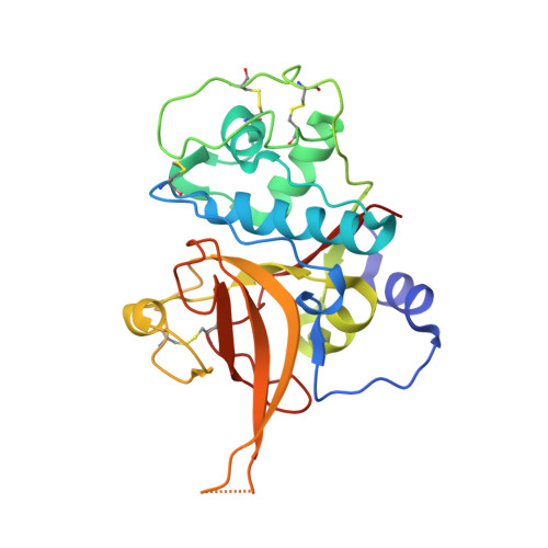
3BPF

Crystal Structure of Falcipain-2 with Its inhibitor, E64


Experimental Data Snapshot

  • Method: X-RAY DIFFRACTION
  • Resolution: 2.90 Å
  • R-Value Free: 0.325 
  • R-Value Work: 0.275 
  • R-Value Observed: 0.278 

Starting Model: experimental
View more details

wwPDB Validation   3D Report Full Report


This is version 1.3 of the entry. See complete history


Literature

Structures of falcipain-2 and falcipain-3 bound to small molecule inhibitors: implications for substrate specificity.

Kerr, I.D.Lee, J.H.Pandey, K.C.Harrison, A.Sajid, M.Rosenthal, P.J.Brinen, L.S.

(2009) J Med Chem 52: 852-857

  • DOI: https://doi.org/10.1021/jm8013663
  • Primary Citation of Related Structures:  
    3BPF, 3BPM

  • PubMed Abstract: 

    Falcipain-2 and falcipain-3 are critical hemoglobinases of Plasmodium falciparum, the most virulent human malaria parasite. We have determined the 2.9 A crystal structure of falcipain-2 in complex with the epoxysuccinate E64 and the 2.5 A crystal structure of falcipain-3 in complex with the aldehyde leupeptin. These complexes represent the first crystal structures of plasmodial cysteine proteases with small molecule inhibitors and the first reported crystal structure of falcipain-3. Our structural analyses indicate that the relative shape and flexibility of the S2 pocket are affected by a number of discrete amino acid substitutions. The cumulative effect of subtle differences, including those at "gatekeeper" positions, may explain the observed kinetic differences between these two closely related enzymes.


  • Organizational Affiliation

    Department of Cellular and Molecular Pharmacology and Department of Pathology, University of California, San Francisco, California 94158, USA.


Macromolecules
Find similar proteins by:  (by identity cutoff)  |  3D Structure
Entity ID: 1
MoleculeChains Sequence LengthOrganismDetailsImage
Cysteine protease falcipain-2
A, B, C, D
241Plasmodium falciparumMutation(s): 0 
EC: 3.4.22
UniProt
Find proteins for Q8I6U4 (Plasmodium falciparum (isolate 3D7))
Explore Q8I6U4 
Go to UniProtKB:  Q8I6U4
Entity Groups  
Sequence Clusters30% Identity50% Identity70% Identity90% Identity95% Identity100% Identity
UniProt GroupQ8I6U4
Sequence Annotations
Expand
  • Reference Sequence
Experimental Data & Validation

Experimental Data

  • Method: X-RAY DIFFRACTION
  • Resolution: 2.90 Å
  • R-Value Free: 0.325 
  • R-Value Work: 0.275 
  • R-Value Observed: 0.278 
  • Space Group: C 2 2 21
Unit Cell:
Length ( Å )Angle ( ˚ )
a = 143.778α = 90
b = 167.81β = 90
c = 177.758γ = 90
Software Package:
Software NamePurpose
REFMACrefinement
ADSCdata collection
MOSFLMdata reduction
XSCALEdata scaling
PHASERphasing

Structure Validation

View Full Validation Report


Entry History 

Deposition Data

Revision History  (Full details and data files)

  • Version 1.0: 2008-12-30
    Type: Initial release
  • Version 1.1: 2011-07-13
    Changes: Advisory, Refinement description, Version format compliance
  • Version 1.2: 2023-08-30
    Changes: Data collection, Database references, Derived calculations, Refinement description
  • Version 1.3: 2024-11-13
    Changes: Structure summary