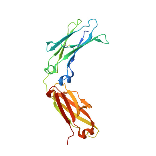
3C2S

Structural Characterization of a Human Fc Fragment Engineered for Lack of Effector Functions


Experimental Data Snapshot

  • Method: X-RAY DIFFRACTION
  • Resolution: 2.30 Å
  • R-Value Free: 0.275 
  • R-Value Work: 0.216 
  • R-Value Observed: 0.219 

Starting Model: experimental
View more details

wwPDB Validation   3D Report Full Report


This is version 2.3 of the entry. See complete history


Literature

Structural characterization of a human Fc fragment engineered for lack of effector functions.

Oganesyan, V.Gao, C.Shirinian, L.Wu, H.Dall'Acqua, W.F.

(2008) Acta Crystallogr D Biol Crystallogr 64: 700-704

  • DOI: https://doi.org/10.1107/S0907444908007877
  • Primary Citation of Related Structures:  
    3C2S

  • PubMed Abstract: 

    The first three-dimensional structure of a human Fc fragment genetically engineered for the elimination of its ability to mediate antibody-dependent cell-mediated cytotoxicity and complement-dependent cytotoxicity is reported. When introduced into the lower hinge and CH2 domain of human IgG1 molecules, the triple mutation L234F/L235E/P331S ('TM') causes a profound decrease in their binding to human CD64, CD32A, CD16 and C1q. Enzymatically produced Fc/TM fragment was crystallized and its structure was solved at a resolution of 2.3 A using molecular replacement. This study revealed that the three-dimensional structure of Fc/TM is very similar to those of other human Fc fragments in the experimentally visible region spanning residues 236-445. Thus, the dramatic broad-ranging effects of TM on IgG binding to several effector molecules cannot be explained in terms of major structural rearrangements in this portion of the Fc.


  • Organizational Affiliation

    Department of Antibody Discovery and Protein Engineering, MedImmune Inc., One MedImmune Way, Gaithersburg, MD 20878, USA.


Macromolecules
Find similar proteins by:  (by identity cutoff)  |  3D Structure
Entity ID: 1
MoleculeChains Sequence LengthOrganismDetailsImage
IGHM protein225Homo sapiensMutation(s): 3 
UniProt & NIH Common Fund Data Resources
Find proteins for P01857 (Homo sapiens)
Explore P01857 
Go to UniProtKB:  P01857
PHAROS:  P01857
GTEx:  ENSG00000211896 
Entity Groups  
Sequence Clusters30% Identity50% Identity70% Identity90% Identity95% Identity100% Identity
UniProt GroupP01857
Glycosylation
Glycosylation Sites: 1Go to GlyGen: P01857-2
Sequence Annotations
Expand
  • Reference Sequence
Oligosaccharides

Help

Entity ID: 2
MoleculeChains Length2D Diagram Glycosylation3D Interactions
beta-D-galactopyranose-(1-4)-2-acetamido-2-deoxy-beta-D-glucopyranose-(1-2)-alpha-D-mannopyranose-(1-6)-[2-acetamido-2-deoxy-beta-D-glucopyranose-(1-2)-alpha-D-mannopyranose-(1-3)]beta-D-mannopyranose-(1-4)-2-acetamido-2-deoxy-beta-D-glucopyranose-(1-4)-[alpha-L-fucopyranose-(1-6)]2-acetamido-2-deoxy-beta-D-glucopyranose
B
9N-Glycosylation
Glycosylation Resources
GlyTouCan:  G27919IH
GlyCosmos:  G27919IH
GlyGen:  G27919IH
Experimental Data & Validation

Experimental Data

  • Method: X-RAY DIFFRACTION
  • Resolution: 2.30 Å
  • R-Value Free: 0.275 
  • R-Value Work: 0.216 
  • R-Value Observed: 0.219 
  • Space Group: C 2 2 21
Unit Cell:
Length ( Å )Angle ( ˚ )
a = 50.178α = 90
b = 147.301β = 90
c = 75.473γ = 90
Software Package:
Software NamePurpose
REFMACrefinement
CrystalCleardata collection
HKL-2000data reduction
SCALEPACKdata scaling
PHASERphasing

Structure Validation

View Full Validation Report


Entry History 

Deposition Data

Revision History  (Full details and data files)

  • Version 1.0: 2008-08-19
    Type: Initial release
  • Version 1.1: 2011-07-13
    Changes: Non-polymer description, Version format compliance
  • Version 2.0: 2020-07-29
    Type: Remediation
    Reason: Carbohydrate remediation
    Changes: Advisory, Atomic model, Data collection, Derived calculations, Structure summary
  • Version 2.1: 2021-10-20
    Changes: Database references, Structure summary
  • Version 2.2: 2023-08-30
    Changes: Data collection, Refinement description
  • Version 2.3: 2024-10-16
    Changes: Structure summary