3CUK | pdb_00003cuk

Crystal structure of human D-amino acid oxidase: bound to an inhibitor


Experimental Data Snapshot

  • Method: X-RAY DIFFRACTION
  • Resolution: 2.49 Å
  • R-Value Free: 
    0.329 (Depositor), 0.224 (DCC) 
  • R-Value Work: 
    0.239 (Depositor), 0.237 (DCC) 
  • R-Value Observed: 
    0.243 (Depositor) 

Starting Model: experimental
View more details

wwPDB Validation 3D Report Full Report

Validation slider image for 3CUK

Ligand Structure Quality Assessment 


This is version 1.3 of the entry. See complete history

Literature

The discovery of fused pyrrole carboxylic acids as novel, potent D-amino acid oxidase (DAO) inhibitors.

Sparey, T.Abeywickrema, P.Almond, S.Brandon, N.Byrne, N.Campbell, A.Hutson, P.H.Jacobson, M.Jones, B.Munshi, S.Pascarella, D.Pike, A.Prasad, G.S.Sachs, N.Sakatis, M.Sardana, V.Venkatraman, S.Young, M.B.

(2008) Bioorg Med Chem Lett 18: 3386-3391

  • DOI: https://doi.org/10.1016/j.bmcl.2008.04.020
  • Primary Citation Related Structures: 
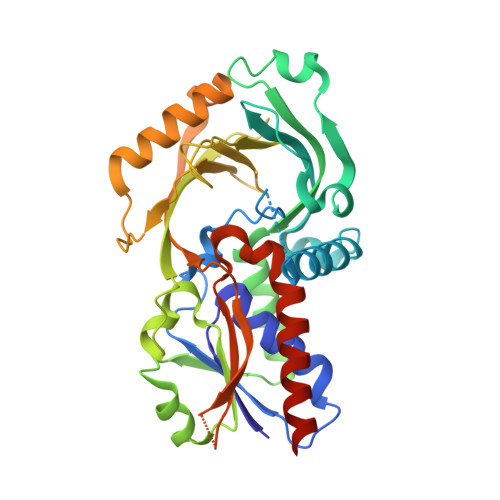
    3CUK

  • PubMed Abstract: 

    The 'NMDA hypofunction hypothesis of schizophrenia' can be tested in a number of ways. DAO is the enzyme primarily responsible for the metabolism of d-serine, a co-agonist for the NMDA receptor. We identified novel DAO inhibitors, in particular, acid 1, which demonstrated moderate potency for DAO in vitro and ex vivo, and raised plasma d-serine levels after dosing ip to rats. In parallel, analogues were prepared to survey the SARs of 1.


  • Organizational Affiliation
    • Department of Medicinal Chemistry, Merck Sharp & Dohme, The Neuroscience Research Centre, Terlings Park, Harlow, Essex CM20 2QR, UK. tim_sparey@merck.com

Macromolecule Content 

  • Total Structure Weight: 161.83 kDa 
  • Atom Count: 10,850 
  • Modeled Residue Count: 1,308 
  • Deposited Residue Count: 1,388 
  • Unique protein chains: 1

Macromolecules

Find similar proteins by:|  3D Structure
Entity ID: 1
MoleculeChains  Sequence LengthOrganismDetailsImage
D-amino-acid oxidase
A, B, C, D
347Homo sapiensMutation(s): 0 
Gene Names: DAODAMOX
EC: 1.4.3.3
UniProt & NIH Common Fund Data Resources
Find proteins for P14920 (Homo sapiens)
Explore P14920 
Go to UniProtKB:  P14920
PHAROS:  P14920
GTEx:  ENSG00000110887 
Entity Groups
Sequence Clusters30% Identity50% Identity70% Identity90% Identity95% Identity100% Identity
UniProt GroupP14920
Sequence Annotations
Expand
Reference Sequence

Small Molecules

Ligands 2 Unique
IDChains Name / Formula / InChI Key2D Diagram3D Interactions
FAD

Query on FAD


Download:Ideal Coordinates CCD File
E [auth A],
G [auth B],
I [auth C],
K [auth D]
FLAVIN-ADENINE DINUCLEOTIDE
C27 H33 N9 O15 P2
VWWQXMAJTJZDQX-UYBVJOGSSA-N
4P5

Query on 4P5


Download:Ideal Coordinates CCD File
F [auth A],
H [auth B],
J [auth C],
L [auth D]
4H-furo[3,2-b]pyrrole-5-carboxylic acid
C7 H5 N O3
MMAIBGHDBYQYDI-UHFFFAOYSA-N

Experimental Data & Validation

Experimental Data

  • Method: X-RAY DIFFRACTION
  • Resolution: 2.49 Å
  • R-Value Free:  0.329 (Depositor), 0.224 (DCC) 
  • R-Value Work:  0.239 (Depositor), 0.237 (DCC) 
  • R-Value Observed: 0.243 (Depositor) 
Space Group: C 1 2 1
Unit Cell:
Length ( Å )Angle ( ˚ )
a = 187.729α = 90
b = 51.145β = 110.45
c = 153.294γ = 90
Software Package:
Software NamePurpose
MOLREPphasing
REFMACrefinement
HKL-2000data scaling

Structure Validation

View Full Validation Report


Ligand Structure Quality Assessment 


Entry History 

Deposition Data

Revision History  (Full details and data files)

  • Version 1.0: 2008-07-22
    Type: Initial release
  • Version 1.1: 2011-07-13
    Changes: Advisory, Version format compliance
  • Version 1.2: 2018-03-14
    Changes: Data collection
  • Version 1.3: 2023-08-30
    Changes: Data collection, Database references, Derived calculations, Refinement description