3CWD | pdb_00003cwd

Molecular recognition of nitro-fatty acids by PPAR gamma


Experimental Data Snapshot

  • Method: X-RAY DIFFRACTION
  • Resolution: 2.40 Å
  • R-Value Free: 
    0.290 (Depositor), 0.302 (DCC) 
  • R-Value Work: 
    0.233 (Depositor), 0.248 (DCC) 
  • R-Value Observed: 
    0.236 (Depositor) 

wwPDB Validation 3D Report Full Report

Validation slider image for 3CWD

Ligand Structure Quality Assessment 


This is version 1.2 of the entry. See complete history

Literature

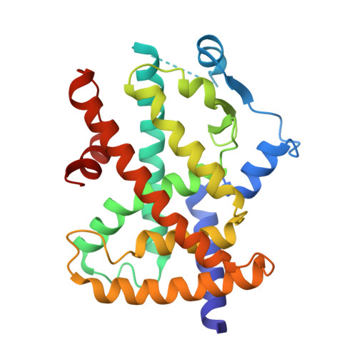
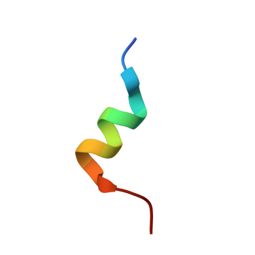
Molecular recognition of nitrated fatty acids by PPAR gamma.

Li, Y.Zhang, J.Schopfer, F.J.Martynowski, D.Garcia-Barrio, M.T.Kovach, A.Suino-Powell, K.Baker, P.R.Freeman, B.A.Chen, Y.E.Xu, H.E.

(2008) Nat Struct Mol Biol 15: 865-867

  • DOI: https://doi.org/10.1038/nsmb.1447
  • Primary Citation Related Structures: 
    3CWD

  • PubMed Abstract: 

    Peroxisome proliferator activated receptor-gamma (PPAR gamma) regulates metabolic homeostasis and adipocyte differentiation, and it is activated by oxidized and nitrated fatty acids. Here we report the crystal structure of the PPAR gamma ligand binding domain bound to nitrated linoleic acid, a potent endogenous ligand of PPAR gamma. Structural and functional studies of receptor-ligand interactions reveal the molecular basis of PPAR gamma discrimination of various naturally occurring fatty acid derivatives.


  • Organizational Affiliation
    • Department of Pharmaceutical Sciences, Center for Pharmacogenetics, 709 Salk Hall, University of Pittsburgh, Pittsburgh, Pennsylvania 15261, USA. yol21@pitt.edu

Macromolecule Content 

  • Total Structure Weight: 66.85 kDa 
  • Atom Count: 4,798 
  • Modeled Residue Count: 549 
  • Deposited Residue Count: 572 
  • Unique protein chains: 2

Macromolecules

Find similar proteins by:|  3D Structure
Entity ID: 1
MoleculeChains  Sequence LengthOrganismDetailsImage
Peroxisome proliferator-activated receptor gamma
A, B
270Homo sapiensMutation(s): 0 
UniProt & NIH Common Fund Data Resources
Find proteins for P37231 (Homo sapiens)
Explore P37231 
Go to UniProtKB:  P37231
PHAROS:  P37231
GTEx:  ENSG00000132170 
Entity Groups
Sequence Clusters30% Identity50% Identity70% Identity90% Identity95% Identity100% Identity
UniProt GroupP37231
Sequence Annotations
Expand
Reference Sequence
Find similar proteins by:  Sequence   |   3D Structure  
Entity ID: 2
MoleculeChains  Sequence LengthOrganismDetailsImage
SRC1-2 PEPTIDE
C, D
16Homo sapiensMutation(s): 0 
Gene Names: NCOA1
EC: 2.3.1.48
UniProt & NIH Common Fund Data Resources
Find proteins for Q15788 (Homo sapiens)
Explore Q15788 
Go to UniProtKB:  Q15788
PHAROS:  Q15788
GTEx:  ENSG00000084676 
Entity Groups
Sequence Clusters30% Identity50% Identity70% Identity90% Identity95% Identity100% Identity
UniProt GroupQ15788
Sequence Annotations
Expand
Reference Sequence

Experimental Data & Validation

Experimental Data

  • Method: X-RAY DIFFRACTION
  • Resolution: 2.40 Å
  • R-Value Free:  0.290 (Depositor), 0.302 (DCC) 
  • R-Value Work:  0.233 (Depositor), 0.248 (DCC) 
  • R-Value Observed: 0.236 (Depositor) 
Space Group: P 21 21 21
Unit Cell:
Length ( Å )Angle ( ˚ )
a = 84.043α = 90
b = 86.197β = 90
c = 97.35γ = 90
Software Package:
Software NamePurpose
REFMACrefinement
HKL-2000data collection
HKL-2000data reduction
HKL-2000data scaling
AMoREphasing

Structure Validation

View Full Validation Report


Ligand Structure Quality Assessment 


Entry History 

Deposition Data

Revision History  (Full details and data files)

  • Version 1.0: 2008-07-08
    Type: Initial release
  • Version 1.1: 2011-07-13
    Changes: Version format compliance
  • Version 1.2: 2024-02-21
    Changes: Data collection, Database references, Derived calculations