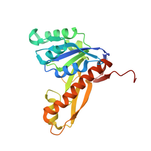
Structures of glycinamide ribonucleotide transformylase (PurN) from Mycobacterium tuberculosis reveal a novel dimer with relevance to drug discovery.
Zhang, Z., Caradoc-Davies, T.T., Dickson, J.M., Baker, E.N., Squire, C.J.(2009) J Mol Biology 389: 722-733
- PubMed: 19394344
- DOI: https://doi.org/10.1016/j.jmb.2009.04.044
- Primary Citation of Related Structures:
3DA8, 3DCJ - PubMed Abstract:
Enzymes from the de novo purine biosynthetic pathway have been exploited for the development of anti-cancer drugs, and represent novel targets for anti-bacterial drug development. In Mycobacterium tuberculosis, the cause of tuberculosis, this pathway has been identified as essential for growth and survival. The structure of M. tuberculosis PurN (MtPurN) has been determined in complex with magnesium and iodide at 1.30 A resolution, and with cofactor analogue, 5-methyltetrahydrofolate (5MTHF) at 2.2 A resolution. The structure shows a Rossmann-type fold that is very similar to the known structures of the human and E. coli PurN proteins. In contrast, MtPurN forms a dimer that is quite different from that formed by the Escherichia coli PurN, and which suggests a mechanism whereby communication could take place between the two active sites. Differences are seen in two active site loops and in the binding mode of the 5MTHF cofactor analogue between the two MtPurN molecules of the dimer. A binding site for halide ions is found in the dimer interface, and bound magnesium and iodide ions in the active site suggest sites that might be exploited in potential drug discovery strategies.
- School of Biological Sciences & Maurice Wilkins Centre for Molecular Biodiscovery, University of Auckland, Private Bag 92010, Auckland 1142, New Zealand.
Organizational Affiliation:


