3DDT | pdb_00003ddt

Crystal structure of the B2 box from MuRF1 in dimeric state


Experimental Data Snapshot

  • Method: X-RAY DIFFRACTION
  • Resolution: 1.90 Å
  • R-Value Free: 
    0.252 (Depositor), 0.253 (DCC) 
  • R-Value Work: 
    0.199 (Depositor), 0.199 (DCC) 
  • R-Value Observed: 
    0.201 (Depositor) 

wwPDB Validation 3D Report Full Report

Validation slider image for 3DDT

This is version 1.2 of the entry. See complete history

Literature

Structural analysis of B-Box 2 from MuRF1: identification of a novel self-association pattern in a RING-like fold

Mrosek, M.Meier, S.Ucurum-Fotiadis, Z.von Castelmur, E.Hedbom, E.Lustig, A.Grzesiek, S.Labeit, D.Labeit, S.Mayans, O.

(2008) Biochemistry 47: 10722-10730

  • DOI: https://doi.org/10.1021/bi800733z
  • Primary Citation Related Structures: 
    3DDT

  • PubMed Abstract: 

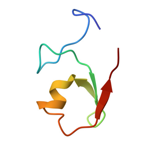
    The B-box motif is the defining feature of the TRIM family of proteins, characterized by a RING finger-B-box-coiled coil tripartite fold. We have elucidated the crystal structure of B-box 2 (B2) from MuRF1, a TRIM protein that supports a wide variety of protein interactions in the sarcomere and regulates the trophic state of striated muscle tissue. MuRF1 B2 coordinates two zinc ions through a cross-brace alpha/beta-topology typical of members of the RING finger superfamily. However, it self-associates into dimers with high affinity. The dimerization pattern is mediated by the helical component of this fold and is unique among RING-like folds. This B2 reveals a long shallow groove that encircles the C-terminal metal binding site ZnII and appears as the defining protein-protein interaction feature of this domain. A cluster of conserved hydrophobic residues in this groove and, in particular, a highly conserved aromatic residue (Y133 in MuRF1 B2) is likely to be central to this role. We expect these findings to aid the future exploration of the cellular function and therapeutic potential of MuRF1.


  • Organizational Affiliation
    • Division of Structural Biology, Biozentrum, University of Basel, Klingelbergstrasse 70, CH-4056 Basel, Switzerland.

Macromolecule Content 

  • Total Structure Weight: 16.22 kDa 
  • Atom Count: 1,215 
  • Modeled Residue Count: 139 
  • Deposited Residue Count: 144 
  • Unique protein chains: 1

Macromolecules

Find similar proteins by:|  3D Structure
Entity ID: 1
MoleculeChains  Sequence LengthOrganismDetailsImage
E3 ubiquitin-protein ligase TRIM63
A, B, C
48Homo sapiensMutation(s): 0 
EC: 6.3.2 (PDB Primary Data), 2.3.2.27 (UniProt)
UniProt & NIH Common Fund Data Resources
Find proteins for Q969Q1 (Homo sapiens)
Explore Q969Q1 
Go to UniProtKB:  Q969Q1
PHAROS:  Q969Q1
GTEx:  ENSG00000158022 
Entity Groups
Sequence Clusters30% Identity50% Identity70% Identity90% Identity95% Identity100% Identity
UniProt GroupQ969Q1
Sequence Annotations
Expand
Reference Sequence

Experimental Data & Validation

Experimental Data

  • Method: X-RAY DIFFRACTION
  • Resolution: 1.90 Å
  • R-Value Free:  0.252 (Depositor), 0.253 (DCC) 
  • R-Value Work:  0.199 (Depositor), 0.199 (DCC) 
  • R-Value Observed: 0.201 (Depositor) 
Space Group: P 65 2 2
Unit Cell:
Length ( Å )Angle ( ˚ )
a = 76.22α = 90
b = 76.22β = 90
c = 146.93γ = 120
Software Package:
Software NamePurpose
PHENIXrefinement
XDSdata reduction
XSCALEdata scaling
SOLVEphasing

Structure Validation

View Full Validation Report


Entry History 

Deposition Data

Revision History  (Full details and data files)

  • Version 1.0: 2008-10-07
    Type: Initial release
  • Version 1.1: 2011-07-13
    Changes: Version format compliance
  • Version 1.2: 2024-03-20
    Changes: Data collection, Database references, Derived calculations