3DPP

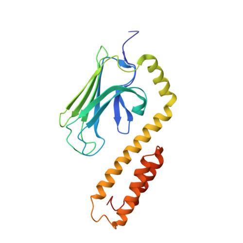
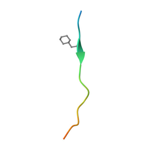
Crystal structure of the substrate binding domain of E. coli DnaK in complex with a long pyrrhocoricin-derived inhibitor peptide (form A)


Experimental Data Snapshot

  • Method: X-RAY DIFFRACTION
  • Resolution: 2.50 Å
  • R-Value Free: 0.316 
  • R-Value Work: 0.237 
  • R-Value Observed: 0.241 

Starting Model: experimental
View more details

wwPDB Validation   3D Report Full Report


This is version 1.4 of the entry. See complete history


Literature

Allosteric coupling between the lid and interdomain linker in DnaK revealed by inhibitor binding studies.

Liebscher, M.Roujeinikova, A.

(2009) J Bacteriol 191: 1456-1462

  • DOI: https://doi.org/10.1128/JB.01131-08
  • Primary Citation of Related Structures:  
    3DPO, 3DPP, 3DPQ

  • PubMed Abstract: 

    The molecular chaperone DnaK assists protein folding and refolding, translocation across membranes, and regulation of the heat shock response. In Escherichia coli, the protein is a target for insect-derived antimicrobial peptides, pyrrhocoricins. We present here the X-ray crystallographic analysis of the E. coli DnaK substrate-binding domain in complex with pyrrhocoricin-derived peptide inhibitors. The structures show that pyrrhocoricins act as site-specific, dual-mode (competitive and allosteric) inhibitors, occupying the substrate-binding tunnel and disrupting the latch between the lid and the beta-sandwich. Our structural analysis revealed an allosteric coupling between the movements of the lid and the interdomain linker, identifying a previously unknown mechanism of the lid-mediated regulation of the chaperone cycle.


  • Organizational Affiliation

    Martin Luther University Halle-Wittenberg, Saale, Germany.


Macromolecules
Find similar proteins by:  (by identity cutoff)  |  3D Structure
Entity ID: 1
MoleculeChains Sequence LengthOrganismDetailsImage
Chaperone protein dnaK
A, B
219Escherichia coli K-12Mutation(s): 0 
Gene Names: dnaKgroPgrpFsegb0014JW0013
UniProt
Find proteins for P0A6Y8 (Escherichia coli (strain K12))
Explore P0A6Y8 
Go to UniProtKB:  P0A6Y8
Entity Groups  
Sequence Clusters30% Identity50% Identity70% Identity90% Identity95% Identity100% Identity
UniProt GroupP0A6Y8
Sequence Annotations
Expand
  • Reference Sequence

Find similar proteins by:  Sequence   |   3D Structure  

Entity ID: 2
MoleculeChains Sequence LengthOrganismDetailsImage
inhibitor peptide
C, D
20N/AMutation(s): 0 
UniProt
Find proteins for P37362 (Pyrrhocoris apterus)
Explore P37362 
Go to UniProtKB:  P37362
Entity Groups  
Sequence Clusters30% Identity50% Identity70% Identity90% Identity95% Identity100% Identity
UniProt GroupP37362
Sequence Annotations
Expand
  • Reference Sequence
Small Molecules
Ligands 1 Unique
IDChains Name / Formula / InChI Key2D Diagram3D Interactions
SO4
Query on SO4

Download Ideal Coordinates CCD File 
E [auth A]SULFATE ION
O4 S
QAOWNCQODCNURD-UHFFFAOYSA-L
Modified Residues  1 Unique
IDChains TypeFormula2D DiagramParent
ALC
Query on ALC
C, D
L-PEPTIDE LINKINGC9 H17 N O2ALA
Experimental Data & Validation

Experimental Data

  • Method: X-RAY DIFFRACTION
  • Resolution: 2.50 Å
  • R-Value Free: 0.316 
  • R-Value Work: 0.237 
  • R-Value Observed: 0.241 
  • Space Group: P 21 21 2
Unit Cell:
Length ( Å )Angle ( ˚ )
a = 77.87α = 90
b = 159.12β = 90
c = 44.79γ = 90
Software Package:
Software NamePurpose
DENZOdata reduction
SCALEPACKdata scaling
REFMACrefinement
PDB_EXTRACTdata extraction
CrystalCleardata collection
d*TREKdata reduction

Structure Validation

View Full Validation Report


Entry History 

Deposition Data

Revision History  (Full details and data files)

  • Version 1.0: 2009-03-10
    Type: Initial release
  • Version 1.1: 2011-07-13
    Changes: Version format compliance
  • Version 1.2: 2017-10-25
    Changes: Refinement description
  • Version 1.3: 2023-08-30
    Changes: Data collection, Database references, Derived calculations, Refinement description
  • Version 1.4: 2024-10-16
    Changes: Structure summary