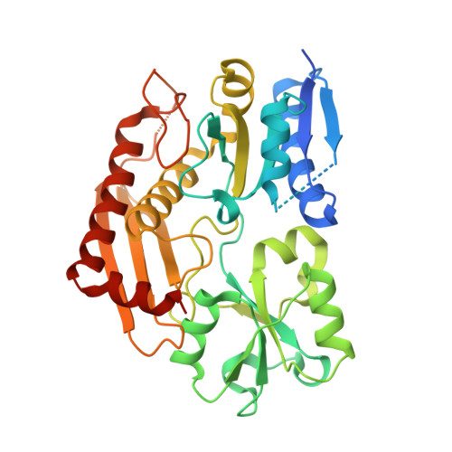
3EQ1

The Crystal Structure of Human Porphobilinogen Deaminase at 2.8A resolution


Experimental Data Snapshot

  • Method: X-RAY DIFFRACTION
  • Resolution: 2.80 Å
  • R-Value Free: 0.289 
  • R-Value Work: 0.259 
  • R-Value Observed: 0.262 

Starting Model: experimental
View more details

wwPDB Validation   3D Report Full Report


Ligand Structure Quality Assessment 


This is version 1.3 of the entry. See complete history


Literature

Structure of human porphobilinogen deaminase at 2.8 A: the molecular basis of acute intermittent porphyria

Gill, R.Kolstoe, S.E.Mohammed, F.Al D-Bass, A.Mosely, J.E.Sarwar, M.Cooper, J.B.Wood, S.P.Shoolingin-Jordan, P.M.

(2009) Biochem J 420: 17-25

  • DOI: https://doi.org/10.1042/BJ20082077
  • Primary Citation of Related Structures:  
    3EQ1

  • PubMed Abstract: 

    Mutations in the human PBGD (porphobilinogen deaminase) gene cause the inherited defect AIP (acute intermittent porphyria). In the present study we report the structure of the human uPBGD (ubiquitous PBGD) mutant, R167Q, that has been determined by X-ray crystallography and refined to 2.8 A (1 A=0.1 nm) resolution (Rfactor=0.26, Rfree=0.29). The protein crystallized in space group P2(1)2(1)2 with two molecules in the asymmetric unit (a=81.0 A, b=104.4 A and c=109.7 A). Phases were obtained by molecular replacement using the Escherichia coli PBGD structure as a search model. The human enzyme is composed of three domains each of approx. 110 amino acids and possesses a dipyrromethane cofactor at the active site, which is located between domains 1 and 2. An ordered sulfate ion is hydrogen-bonded to Arg26 and Ser28 at the proposed substrate-binding site in domain 1. An insert of 29 amino acid residues, present only in mammalian PBGD enzymes, has been modelled into domain 3 where it extends helix alpha2(3) and forms a beta-hairpin structure that contributes to a continuous hydrogen-bonding network spanning domains 1 and 3. The structural and functional implications of the R167Q mutation and other mutations that result in AIP are discussed.


  • Organizational Affiliation

    Centre for Amyloidosis and Acute Phase Proteins, Department of Medicine, UCL Medical School, Rowland Hill Street, London NW3 2PF, UK.


Macromolecules
Find similar proteins by:  (by identity cutoff)  |  3D Structure
Entity ID: 1
MoleculeChains Sequence LengthOrganismDetailsImage
Porphobilinogen deaminase
A, B
361Homo sapiensMutation(s): 1 
EC: 2.5.1.61
UniProt & NIH Common Fund Data Resources
Find proteins for P08397 (Homo sapiens)
Explore P08397 
Go to UniProtKB:  P08397
PHAROS:  P08397
GTEx:  ENSG00000256269 
Entity Groups  
Sequence Clusters30% Identity50% Identity70% Identity90% Identity95% Identity100% Identity
UniProt GroupP08397
Sequence Annotations
Expand
  • Reference Sequence
Small Molecules
Ligands 2 Unique
IDChains Name / Formula / InChI Key2D Diagram3D Interactions
DPM
Query on DPM

Download Ideal Coordinates CCD File 
C [auth A],
G [auth B]
3-[5-{[3-(2-carboxyethyl)-4-(carboxymethyl)-5-methyl-1H-pyrrol-2-yl]methyl}-4-(carboxymethyl)-1H-pyrrol-3-yl]propanoic acid
C20 H24 N2 O8
LCAXMKQKEYTFDM-UHFFFAOYSA-N
SO4
Query on SO4

Download Ideal Coordinates CCD File 
D [auth A]
E [auth A]
F [auth A]
H [auth B]
I [auth B]
D [auth A],
E [auth A],
F [auth A],
H [auth B],
I [auth B],
J [auth B],
K [auth B]
SULFATE ION
O4 S
QAOWNCQODCNURD-UHFFFAOYSA-L
Experimental Data & Validation

Experimental Data

  • Method: X-RAY DIFFRACTION
  • Resolution: 2.80 Å
  • R-Value Free: 0.289 
  • R-Value Work: 0.259 
  • R-Value Observed: 0.262 
  • Space Group: P 21 21 2
Unit Cell:
Length ( Å )Angle ( ˚ )
a = 81.083α = 90
b = 104.435β = 90
c = 109.732γ = 90
Software Package:
Software NamePurpose
MOLREPphasing
PHENIXrefinement
MOSFLMdata reduction
SCALAdata scaling

Structure Validation

View Full Validation Report


Ligand Structure Quality Assessment 


Entry History 

Deposition Data

Revision History  (Full details and data files)

  • Version 1.0: 2009-03-03
    Type: Initial release
  • Version 1.1: 2011-07-13
    Changes: Version format compliance
  • Version 1.2: 2021-11-10
    Changes: Database references, Derived calculations
  • Version 1.3: 2023-11-01
    Changes: Data collection, Refinement description