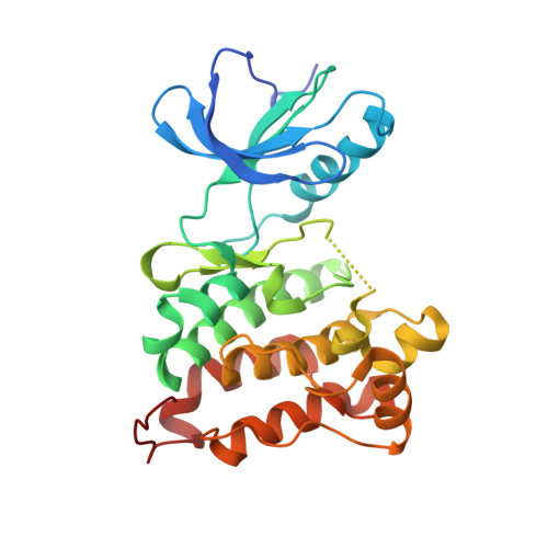
3F3V

Kinase domain of cSrc in complex with inhibitor RL45 (Type II)


Experimental Data Snapshot

  • Method: X-RAY DIFFRACTION
  • Resolution: 2.60 Å
  • R-Value Free: 0.284 
  • R-Value Work: 0.210 
  • R-Value Observed: 0.213 

Starting Model: experimental
View more details

wwPDB Validation   3D Report Full Report


Ligand Structure Quality Assessment 


This is version 1.3 of the entry. See complete history


Literature

Hybrid compound design to overcome the gatekeeper T338M mutation in cSrc

Getlik, M.Grutter, C.Simard, J.R.Kluter, S.Rabiller, M.Rode, H.B.Robubi, A.Rauh, D.

(2009) J Med Chem 52: 3915-3926

  • DOI: https://doi.org/10.1021/jm9002928
  • Primary Citation of Related Structures:  
    3F3V, 3F3W, 3G5D

  • PubMed Abstract: 

    The emergence of drug resistance remains a fundamental challenge in the development of kinase inhibitors that are effective over long-term treatments. Allosteric inhibitors that bind to sites lying outside the highly conserved ATP pocket are thought to be more selective than ATP-competitive inhibitors and may circumvent some mechanisms of drug resistance. Crystal structures of type I and allosteric type III inhibitors in complex with the tyrosine kinase cSrc allowed us to employ principles of structure-based design to develop these scaffolds into potent type II kinase inhibitors. One of these compounds, 3c (RL46), disrupts FAK-mediated focal adhesions in cancer cells via direct inhibition of cSrc. Details gleaned from crystal structures revealed a key feature of a subset of these compounds, a surprising flexibility in the vicinity of the gatekeeper residue that allows these compounds to overcome a dasatinib-resistant gatekeeper mutation emerging in cSrc.


  • Organizational Affiliation

    Chemical Genomics Centre of the Max Planck Society, D-44227 Dortmund, Germany.


Macromolecules
Find similar proteins by:  (by identity cutoff)  |  3D Structure
Entity ID: 1
MoleculeChains Sequence LengthOrganismDetailsImage
Proto-oncogene tyrosine-protein kinase Src
A, B
286Gallus gallusMutation(s): 1 
EC: 2.7.10.2
UniProt
Find proteins for P00523 (Gallus gallus)
Explore P00523 
Go to UniProtKB:  P00523
Entity Groups  
Sequence Clusters30% Identity50% Identity70% Identity90% Identity95% Identity100% Identity
UniProt GroupP00523
Sequence Annotations
Expand
  • Reference Sequence
Small Molecules
Ligands 1 Unique
IDChains Name / Formula / InChI Key2D Diagram3D Interactions
1BU
Query on 1BU

Download Ideal Coordinates CCD File 
C [auth A],
D [auth B]
1-{4-[(6-aminoquinazolin-4-yl)amino]phenyl}-3-[3-tert-butyl-1-(3-methylphenyl)-1H-pyrazol-5-yl]urea
C29 H30 N8 O
ZKESOLNZMJUNTF-UHFFFAOYSA-N
Binding Affinity Annotations 
IDSourceBinding Affinity
1BU BindingDB:  3F3V Kd: 73 (nM) from 1 assay(s)
IC50: min: 21, max: 34 (nM) from 2 assay(s)
PDBBind:  3F3V Kd: 73 (nM) from 1 assay(s)
Experimental Data & Validation

Experimental Data

  • Method: X-RAY DIFFRACTION
  • Resolution: 2.60 Å
  • R-Value Free: 0.284 
  • R-Value Work: 0.210 
  • R-Value Observed: 0.213 
  • Space Group: P 1
Unit Cell:
Length ( Å )Angle ( ˚ )
a = 42.055α = 79.06
b = 63.397β = 87.72
c = 74.955γ = 90.08
Software Package:
Software NamePurpose
XSCALEdata scaling
PHASERphasing
REFMACrefinement
PDB_EXTRACTdata extraction
XDSdata scaling

Structure Validation

View Full Validation Report


Ligand Structure Quality Assessment 


Entry History 

Deposition Data

Revision History  (Full details and data files)

  • Version 1.0: 2009-06-02
    Type: Initial release
  • Version 1.1: 2011-07-13
    Changes: Advisory, Version format compliance
  • Version 1.2: 2021-11-10
    Changes: Database references, Derived calculations
  • Version 1.3: 2023-11-01
    Changes: Data collection, Refinement description