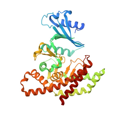
3G15

Crystal structure of human choline kinase alpha in complex with hemicholinium-3 and ADP


Experimental Data Snapshot

  • Method: X-RAY DIFFRACTION
  • Resolution: 1.70 Å
  • R-Value Free: 0.257 
  • R-Value Work: 0.220 
  • R-Value Observed: 0.221 

Starting Model: experimental
View more details

wwPDB Validation   3D Report Full Report


Ligand Structure Quality Assessment 


This is version 1.4 of the entry. See complete history


Literature

Crystal structures of human choline kinase isoforms in complex with hemicholinium-3: single amino acid near the active site influences inhibitor sensitivity.

Hong, B.S.Allali-Hassani, A.Tempel, W.Finerty, P.J.Mackenzie, F.Dimov, S.Vedadi, M.Park, H.W.

(2010) J Biol Chem 285: 16330-16340

  • DOI: https://doi.org/10.1074/jbc.M109.039024
  • Primary Citation of Related Structures:  
    3FEG, 3G15, 3LQ3

  • PubMed Abstract: 

    Human choline kinase (ChoK) catalyzes the first reaction in phosphatidylcholine biosynthesis and exists as ChoKalpha (alpha1 and alpha2) and ChoKbeta isoforms. Recent studies suggest that ChoK is implicated in tumorigenesis and emerging as an attractive target for anticancer chemotherapy. To extend our understanding of the molecular mechanism of ChoK inhibition, we have determined the high resolution x-ray structures of the ChoKalpha1 and ChoKbeta isoforms in complex with hemicholinium-3 (HC-3), a known inhibitor of ChoK. In both structures, HC-3 bound at the conserved hydrophobic groove on the C-terminal lobe. One of the HC-3 oxazinium rings complexed with ChoKalpha1 occupied the choline-binding pocket, providing a structural explanation for its inhibitory action. Interestingly, the HC-3 molecule co-crystallized with ChoKbeta was phosphorylated in the choline binding site. This phosphorylation, albeit occurring at a very slow rate, was confirmed experimentally by mass spectroscopy and radioactive assays. Detailed kinetic studies revealed that HC-3 is a much more potent inhibitor for ChoKalpha isoforms (alpha1 and alpha2) compared with ChoKbeta. Mutational studies based on the structures of both inhibitor-bound ChoK complexes demonstrated that Leu-401 of ChoKalpha2 (equivalent to Leu-419 of ChoKalpha1), or the corresponding residue Phe-352 of ChoKbeta, which is one of the hydrophobic residues neighboring the active site, influences the plasticity of the HC-3-binding groove, thereby playing a key role in HC-3 sensitivity and phosphorylation.


  • Organizational Affiliation

    Structural Genomics Consortium, University of Toronto, Toronto, Ontario M5G 1L7, Canada.


Macromolecules
Find similar proteins by:  (by identity cutoff)  |  3D Structure
Entity ID: 1
MoleculeChains Sequence LengthOrganismDetailsImage
Choline kinase alpha
A, B
401Homo sapiensMutation(s): 3 
Gene Names: CHKACHKCKI
EC: 2.7.1.32 (PDB Primary Data), 2.7.1.82 (UniProt)
UniProt & NIH Common Fund Data Resources
Find proteins for P35790 (Homo sapiens)
Explore P35790 
Go to UniProtKB:  P35790
PHAROS:  P35790
GTEx:  ENSG00000110721 
Entity Groups  
Sequence Clusters30% Identity50% Identity70% Identity90% Identity95% Identity100% Identity
UniProt GroupP35790
Sequence Annotations
Expand
  • Reference Sequence
Small Molecules
Ligands 5 Unique
IDChains Name / Formula / InChI Key2D Diagram3D Interactions
ADP
Query on ADP

Download Ideal Coordinates CCD File 
C [auth A],
T [auth B]
ADENOSINE-5'-DIPHOSPHATE
C10 H15 N5 O10 P2
XTWYTFMLZFPYCI-KQYNXXCUSA-N
HC6
Query on HC6

Download Ideal Coordinates CCD File 
E [auth A],
V [auth B]
(2S,2'S)-2,2'-biphenyl-4,4'-diylbis(2-hydroxy-4,4-dimethylmorpholin-4-ium)
C24 H34 N2 O4
JIWUESGGKYLPPG-DNQXCXABSA-N
SO4
Query on SO4

Download Ideal Coordinates CCD File 
F [auth A],
W [auth B]
SULFATE ION
O4 S
QAOWNCQODCNURD-UHFFFAOYSA-L
MG
Query on MG

Download Ideal Coordinates CCD File 
D [auth A],
G [auth A],
U [auth B],
X [auth B]
MAGNESIUM ION
Mg
JLVVSXFLKOJNIY-UHFFFAOYSA-N
UNX
Query on UNX

Download Ideal Coordinates CCD File 
AA [auth B]
BA [auth B]
CA [auth B]
DA [auth B]
EA [auth B]
AA [auth B],
BA [auth B],
CA [auth B],
DA [auth B],
EA [auth B],
FA [auth B],
GA [auth B],
H [auth A],
I [auth A],
J [auth A],
K [auth A],
L [auth A],
M [auth A],
N [auth A],
O [auth A],
P [auth A],
Q [auth A],
R [auth A],
S [auth A],
Y [auth B],
Z [auth B]
UNKNOWN ATOM OR ION
X
Binding Affinity Annotations 
IDSourceBinding Affinity
HC6 PDBBind:  3G15 Kd: 100 (nM) from 1 assay(s)
Experimental Data & Validation

Experimental Data

  • Method: X-RAY DIFFRACTION
  • Resolution: 1.70 Å
  • R-Value Free: 0.257 
  • R-Value Work: 0.220 
  • R-Value Observed: 0.221 
  • Space Group: P 21 21 21
Unit Cell:
Length ( Å )Angle ( ˚ )
a = 55.802α = 90
b = 118.986β = 90
c = 131.039γ = 90
Software Package:
Software NamePurpose
DENZOdata reduction
SCALEPACKdata scaling
PHASERphasing
REFMACrefinement
PDB_EXTRACTdata extraction
Cootmodel building
MolProbitymodel building
ARP/wARPmodel building

Structure Validation

View Full Validation Report


Ligand Structure Quality Assessment 


Entry History 

Deposition Data

Revision History  (Full details and data files)

  • Version 1.0: 2009-02-10
    Type: Initial release
  • Version 1.1: 2011-07-13
    Changes: Version format compliance
  • Version 1.2: 2017-11-01
    Changes: Refinement description
  • Version 1.3: 2020-10-21
    Changes: Data collection, Database references, Derived calculations
  • Version 1.4: 2023-09-06
    Changes: Data collection, Database references, Refinement description