3GLM | pdb_00003glm

Glutaconyl-coA decarboxylase A subunit from Clostridium symbiosum co-crystallized with crotonyl-coA


Experimental Data Snapshot

  • Method: X-RAY DIFFRACTION
  • Resolution: 2.50 Å
  • R-Value Free: 
    0.230 (Depositor), 0.185 (DCC) 
  • R-Value Work: 
    0.193 (Depositor) 
  • R-Value Observed: 
    0.194 (Depositor) 

wwPDB Validation 3D Report Full Report

Validation slider image for 3GLM

Ligand Structure Quality Assessment 


This is version 1.3 of the entry. See complete history

Literature

An asymmetric model for Na+-translocating glutaconyl-CoA decarboxylases

Kress, D.Brugel, D.Schall, I.Linder, D.Buckel, W.Essen, L.-O.

(2009) J Biological Chem 284: 28401-28409

  • DOI: https://doi.org/10.1074/jbc.M109.037762
  • Primary Citation Related Structures: 
    3GF3, 3GF7, 3GLM, 3GMA

  • PubMed Abstract: 

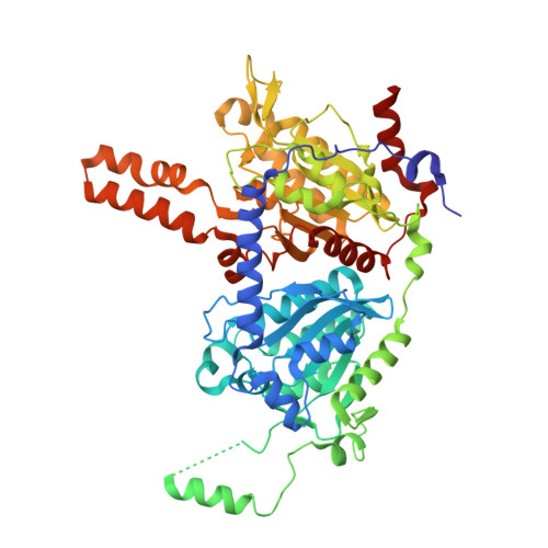
    Glutaconyl-CoA decarboxylase (Gcd) couples the biotin-dependent decarboxylation of glutaconyl-CoA with the generation of an electrochemical Na(+) gradient. Sequencing of the genes encoding all subunits of the Clostridium symbiosum decarboxylase membrane complex revealed that it comprises two distinct biotin carrier subunits, GcdC(1) and GcdC(2), which differ in the length of a central alanine- and proline-rich linker domain. Co-crystallization of the decarboxylase subunit GcdA with the substrate glutaconyl-CoA, the product crotonyl-CoA, and the substrate analogue glutaryl-CoA, respectively, resulted in a high resolution model for substrate binding and catalysis revealing remarkable structural changes upon substrate binding. Unlike the GcdA structure from Acidaminococcus fermentans, these data suggest that in intact Gcd complexes, GcdA is associated as a tetramer crisscrossed by a network of solvent-filled tunnels.


  • Organizational Affiliation
    • Biochemie, Fachbereich Chemie, Philipps-Universität Marburg, D-35032 Marburg, Germany.

Macromolecule Content 

  • Total Structure Weight: 263.58 kDa 
  • Atom Count: 18,142 
  • Modeled Residue Count: 2,244 
  • Deposited Residue Count: 2,352 
  • Unique protein chains: 1

Macromolecules

Find similar proteins by:|  3D Structure
Entity ID: 1
MoleculeChains  Sequence LengthOrganismDetailsImage
Glutaconyl-CoA decarboxylase subunit A
A, B, C, D
588[Clostridium] symbiosumMutation(s): 0 
Gene Names: gcdA
EC: 4.1.1.70
UniProt
Find proteins for B7TVP1 (Clostridium symbiosum)
Explore B7TVP1 
Go to UniProtKB:  B7TVP1
Entity Groups
Sequence Clusters30% Identity50% Identity70% Identity90% Identity95% Identity100% Identity
UniProt GroupB7TVP1
Sequence Annotations
Expand
Reference Sequence

Small Molecules

Ligands 2 Unique
IDChains Name / Formula / InChI Key2D Diagram3D Interactions
COO

Query on COO


Download:Ideal Coordinates CCD File
F [auth A],
H [auth B],
J [auth C],
L [auth D]
CROTONYL COENZYME A
C25 H40 N7 O17 P3 S
KFWWCMJSYSSPSK-XBTRWLRFSA-N
CL

Query on CL


Download:Ideal Coordinates CCD File
E [auth A],
G [auth B],
I [auth C],
K [auth D]
CHLORIDE ION
Cl
VEXZGXHMUGYJMC-UHFFFAOYSA-M

Experimental Data & Validation

Experimental Data

  • Method: X-RAY DIFFRACTION
  • Resolution: 2.50 Å
  • R-Value Free:  0.230 (Depositor), 0.185 (DCC) 
  • R-Value Work:  0.193 (Depositor) 
  • R-Value Observed: 0.194 (Depositor) 
Space Group: C 1 2 1
Unit Cell:
Length ( Å )Angle ( ˚ )
a = 101.5α = 90
b = 167.45β = 89.98
c = 138.11γ = 90
Software Package:
Software NamePurpose
MOSFLMdata reduction
SCALAdata scaling
REFMACrefinement
PDB_EXTRACTdata extraction

Structure Validation

View Full Validation Report


Ligand Structure Quality Assessment 


Entry History 

Deposition Data

Revision History  (Full details and data files)

  • Version 1.0: 2009-07-28
    Type: Initial release
  • Version 1.1: 2011-07-13
    Changes: Version format compliance
  • Version 1.2: 2014-02-05
    Changes: Database references
  • Version 1.3: 2024-03-20
    Changes: Data collection, Database references, Derived calculations, Refinement description