3HDN | pdb_00003hdn

Crystal structure of serum and glucocorticoid-regulated kinase 1 in complex with compound 2


Experimental Data Snapshot

  • Method: X-RAY DIFFRACTION
  • Resolution: 3.10 Å
  • R-Value Free: 
    0.278 (Depositor), 0.270 (DCC) 
  • R-Value Work: 
    0.250 (Depositor), 0.242 (DCC) 
  • R-Value Observed: 
    0.251 (Depositor) 

wwPDB Validation 3D Report Full Report

Validation slider image for 3HDN

Ligand Structure Quality Assessment 


This is version 1.3 of the entry. See complete history

Literature

Design and synthesis of orally bioavailable serum and glucocorticoid-regulated kinase 1 (SGK1) inhibitors.

Hammond, M.Washburn, D.G.Hoang, H.T.Manns, S.Frazee, J.S.Nakamura, H.Patterson, J.R.Trizna, W.Wu, C.Azzarano, L.M.Nagilla, R.Nord, M.Trejo, R.Head, M.S.Zhao, B.Smallwood, A.M.Hightower, K.Laping, N.J.Schnackenberg, C.G.Thompson, S.K.

(2009) Bioorg Med Chem Lett 19: 4441-4445

  • DOI: https://doi.org/10.1016/j.bmcl.2009.05.051
  • Primary Citation Related Structures: 
    3HDM, 3HDN

  • PubMed Abstract: 

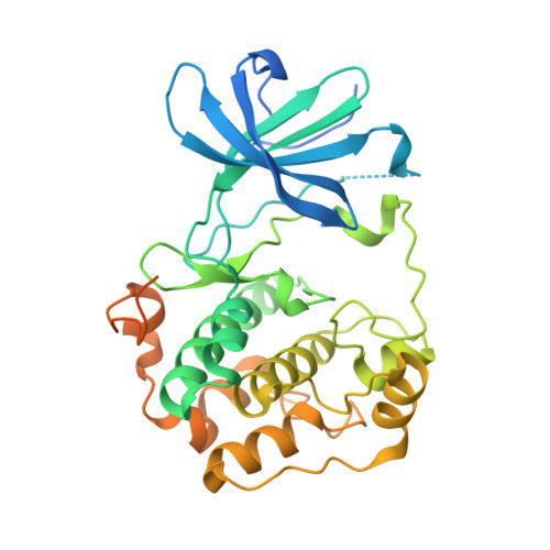
    The lead serum and glucocorticoid-related kinase 1 (SGK1) inhibitors 4-(5-phenyl-1H-pyrrolo[2,3-b]pyridin-3-yl)benzoic acid (1) and {4-[5-(2-naphthalenyl)-1H-pyrrolo[2,3-b]pyridin-3-yl]phenyl}acetic acid (2) suffer from low DNAUC values in rat, due in part to formation and excretion of glucuronic acid conjugates. These PK/glucuronidation issues were addressed either by incorporating a substituent on the 3-phenyl ring ortho to the key carboxylate functionality of 1 or by substituting on the group in between the carboxylate and phenyl ring of 2. Three of these analogs have been identified as having good SGK1 inhibition potency and have DNAUC values suitable for in vivo testing.


  • Organizational Affiliation
    • Department of Chemistry, Metabolic Pathways Centre for Excellence in Drug Discovery, GlaxoSmithKline Pharmaceuticals, 709 Swedeland Road, King of Prussia, PA 19406, USA. marlys.2.hammond@gsk.com

Macromolecule Content 

  • Total Structure Weight: 42.68 kDa 
  • Atom Count: 2,271 
  • Modeled Residue Count: 280 
  • Deposited Residue Count: 373 
  • Unique protein chains: 1

Macromolecules

Find similar proteins by:|  3D Structure
Entity ID: 1
MoleculeChains  Sequence LengthOrganismDetailsImage
Serine/threonine-protein kinase Sgk1373Homo sapiensMutation(s): 4 
Gene Names: SGKSGK1
EC: 2.7.11.1
UniProt & NIH Common Fund Data Resources
Find proteins for O00141 (Homo sapiens)
Explore O00141 
Go to UniProtKB:  O00141
PHAROS:  O00141
GTEx:  ENSG00000118515 
Entity Groups
Sequence Clusters30% Identity50% Identity70% Identity90% Identity95% Identity100% Identity
UniProt GroupO00141
Sequence Annotations
Expand
Reference Sequence

Small Molecules

Ligands 1 Unique
IDChains Name / Formula / InChI Key2D Diagram3D Interactions
GMG

Query on GMG


Download:Ideal Coordinates CCD File
B [auth A][4-(5-naphthalen-2-yl-1H-pyrrolo[2,3-b]pyridin-3-yl)phenyl]acetic acid
C25 H18 N2 O2
SWXKLXXVFMYMDP-UHFFFAOYSA-N

Experimental Data & Validation

Experimental Data

  • Method: X-RAY DIFFRACTION
  • Resolution: 3.10 Å
  • R-Value Free:  0.278 (Depositor), 0.270 (DCC) 
  • R-Value Work:  0.250 (Depositor), 0.242 (DCC) 
  • R-Value Observed: 0.251 (Depositor) 
Space Group: P 65 2 2
Unit Cell:
Length ( Å )Angle ( ˚ )
a = 96.812α = 90
b = 96.812β = 90
c = 150.779γ = 120
Software Package:
Software NamePurpose
REFMACrefinement

Structure Validation

View Full Validation Report


Ligand Structure Quality Assessment 


Entry History 

Deposition Data

Revision History  (Full details and data files)

  • Version 1.0: 2009-06-30
    Type: Initial release
  • Version 1.1: 2011-07-13
    Changes: Advisory, Version format compliance
  • Version 1.2: 2021-10-13
    Changes: Database references, Derived calculations
  • Version 1.3: 2024-02-21
    Changes: Data collection