3HG5 | pdb_00003hg5

Human alpha-galactosidase catalytic mechanism 4. Product bound


Experimental Data Snapshot

  • Method: X-RAY DIFFRACTION
  • Resolution: 2.30 Å
  • R-Value Free: 
    0.227 (Depositor), 0.225 (DCC) 
  • R-Value Work: 
    0.191 (Depositor), 0.189 (DCC) 
  • R-Value Observed: 
    0.192 (Depositor) 

Starting Model: experimental
View more details

wwPDB Validation 3D Report Full Report

Validation slider image for 3HG5

Ligand Structure Quality Assessment 


This is version 2.2 of the entry. See complete history

Literature

Catalytic mechanism of human alpha-galactosidase.

Guce, A.I.Clark, N.E.Salgado, E.N.Ivanen, D.R.Kulminskaya, A.A.Brumer, H.Garman, S.C.

(2010) J Biological Chem 285: 3625-3632

  • DOI: https://doi.org/10.1074/jbc.M109.060145
  • Primary Citation Related Structures: 
    3HG2, 3HG3, 3HG4, 3HG5

  • PubMed Abstract: 

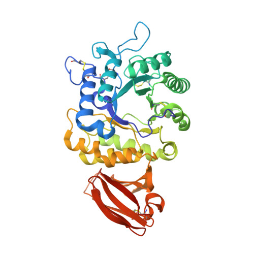
    The enzyme alpha-galactosidase (alpha-GAL, also known as alpha-GAL A; E.C. 3.2.1.22) is responsible for the breakdown of alpha-galactosides in the lysosome. Defects in human alpha-GAL lead to the development of Fabry disease, a lysosomal storage disorder characterized by the buildup of alpha-galactosylated substrates in the tissues. alpha-GAL is an active target of clinical research: there are currently two treatment options for Fabry disease, recombinant enzyme replacement therapy (approved in the United States in 2003) and pharmacological chaperone therapy (currently in clinical trials). Previously, we have reported the structure of human alpha-GAL, which revealed the overall structure of the enzyme and established the locations of hundreds of mutations that lead to the development of Fabry disease. Here, we describe the catalytic mechanism of the enzyme derived from x-ray crystal structures of each of the four stages of the double displacement reaction mechanism. Use of a difluoro-alpha-galactopyranoside allowed trapping of a covalent intermediate. The ensemble of structures reveals distortion of the ligand into a (1)S(3) skew (or twist) boat conformation in the middle of the reaction cycle. The high resolution structures of each step in the catalytic cycle will allow for improved drug design efforts on alpha-GAL and other glycoside hydrolase family 27 enzymes by developing ligands that specifically target different states of the catalytic cycle. Additionally, the structures revealed a second ligand-binding site suitable for targeting by novel pharmacological chaperones.


  • Organizational Affiliation
    • Departments of Chemistry, University of Massachusetts, Amherst, Massachusetts 01003.

Macromolecule Content 

  • Total Structure Weight: 93.95 kDa 
  • Atom Count: 6,692 
  • Modeled Residue Count: 781 
  • Deposited Residue Count: 796 
  • Unique protein chains: 1

Macromolecules

Find similar proteins by:|  3D Structure
Entity ID: 1
MoleculeChains  Sequence LengthOrganismDetailsImage
Alpha-galactosidase A
A, B
398Homo sapiensMutation(s): 0 
Gene Names: GLA
EC: 3.2.1.22
UniProt & NIH Common Fund Data Resources
Find proteins for P06280 (Homo sapiens)
Explore P06280 
Go to UniProtKB:  P06280
PHAROS:  P06280
GTEx:  ENSG00000102393 
Entity Groups
Sequence Clusters30% Identity50% Identity70% Identity90% Identity95% Identity100% Identity
UniProt GroupP06280
Glycosylation
Glycosylation Sites: 3Go to GlyGen: P06280-1
Sequence Annotations
Expand
Reference Sequence

Oligosaccharides

Help  
Entity ID: 2
MoleculeChains Length2D Diagram GlycosylationD Interactions
beta-D-mannopyranose-(1-4)-2-acetamido-2-deoxy-beta-D-glucopyranose-(1-4)-2-acetamido-2-deoxy-beta-D-glucopyranose
C
3N-Glycosylation
Glycosylation Resources
GlyTouCan: G15407YE
GlyCosmos: G15407YE
GlyGen: G15407YE
Entity ID: 3
MoleculeChains Length2D Diagram GlycosylationD Interactions
alpha-D-mannopyranose-(1-3)-beta-D-mannopyranose-(1-4)-2-acetamido-2-deoxy-beta-D-glucopyranose-(1-4)-2-acetamido-2-deoxy-beta-D-glucopyranose
D
4N-Glycosylation
Glycosylation Resources
GlyTouCan: G81315DD
GlyCosmos: G81315DD
GlyGen: G81315DD

Small Molecules

Ligands 5 Unique
IDChains Name / Formula / InChI Key2D Diagram3D Interactions
NAG

Query on NAG


Download:Ideal Coordinates CCD File
E [auth A],
F [auth A],
L [auth B]
2-acetamido-2-deoxy-beta-D-glucopyranose
C8 H15 N O6
OVRNDRQMDRJTHS-FMDGEEDCSA-N
GAL

Query on GAL


Download:Ideal Coordinates CCD File
N [auth B]beta-D-galactopyranose
C6 H12 O6
WQZGKKKJIJFFOK-FPRJBGLDSA-N
GLA

Query on GLA


Download:Ideal Coordinates CCD File
G [auth A],
M [auth B]
alpha-D-galactopyranose
C6 H12 O6
WQZGKKKJIJFFOK-PHYPRBDBSA-N
SO4

Query on SO4


Download:Ideal Coordinates CCD File
H [auth A],
I [auth A],
O [auth B],
P [auth B]
SULFATE ION
O4 S
QAOWNCQODCNURD-UHFFFAOYSA-L
ACY

Query on ACY


Download:Ideal Coordinates CCD File
J [auth A],
K [auth A],
Q [auth B],
R [auth B]
ACETIC ACID
C2 H4 O2
QTBSBXVTEAMEQO-UHFFFAOYSA-N

Experimental Data & Validation

Experimental Data

  • Method: X-RAY DIFFRACTION
  • Resolution: 2.30 Å
  • R-Value Free:  0.227 (Depositor), 0.225 (DCC) 
  • R-Value Work:  0.191 (Depositor), 0.189 (DCC) 
  • R-Value Observed: 0.192 (Depositor) 
Space Group: P 32 2 1
Unit Cell:
Length ( Å )Angle ( ˚ )
a = 90.813α = 90
b = 90.813β = 90
c = 217.166γ = 120
Software Package:
Software NamePurpose
DENZOdata reduction
SCALEPACKdata scaling
REFMACrefinement
PDB_EXTRACTdata extraction
HKL-2000data collection
HKL-2000data reduction
HKL-2000data scaling

Structure Validation

View Full Validation Report


Ligand Structure Quality Assessment 


Entry History 

Deposition Data

Revision History  (Full details and data files)

  • Version 1.0: 2009-11-24
    Type: Initial release
  • Version 1.1: 2011-07-13
    Changes: Advisory, Version format compliance
  • Version 1.2: 2017-11-01
    Changes: Refinement description
  • Version 2.0: 2020-07-29
    Type: Remediation
    Reason: Carbohydrate remediation
    Changes: Atomic model, Data collection, Derived calculations, Structure summary
  • Version 2.1: 2024-04-03
    Changes: Data collection, Database references, Refinement description, Structure summary
  • Version 2.2: 2024-11-06
    Changes: Structure summary