3I2I | pdb_00003i2i

Cocaine Esterase with mutation T172R, bound to DTT adduct


Experimental Data Snapshot

  • Method: X-RAY DIFFRACTION
  • Resolution: 2.14 Å
  • R-Value Free: 
    0.180 (Depositor), 0.158 (DCC) 
  • R-Value Work: 
    0.159 (Depositor) 
  • R-Value Observed: 
    0.159 (Depositor) 

Starting Model: experimental
View more details

wwPDB Validation 3D Report Full Report

Validation slider image for 3I2I

Ligand Structure Quality Assessment 


This is version 1.5 of the entry. See complete history

Literature

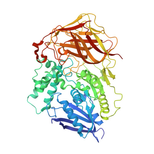
Structural analysis of thermostabilizing mutations of cocaine esterase.

Narasimhan, D.Nance, M.R.Gao, D.Ko, M.C.Macdonald, J.Tamburi, P.Yoon, D.Landry, D.M.Woods, J.H.Zhan, C.G.Tesmer, J.J.Sunahara, R.K.

(2010) Protein Eng Des Sel 23: 537-547

  • DOI: https://doi.org/10.1093/protein/gzq025
  • Primary Citation Related Structures: 
    3I2F, 3I2G, 3I2H, 3I2I, 3I2J, 3I2K

  • PubMed Abstract: 

    Cocaine is considered to be the most addictive of all substances of abuse and mediates its effects by inhibiting monoamine transporters, primarily the dopamine transporters. There are currently no small molecules that can be used to combat its toxic and addictive properties, in part because of the difficulty of developing compounds that inhibit cocaine binding without having intrinsic effects on dopamine transport. Most of the effective cocaine inhibitors also display addictive properties. We have recently reported the use of cocaine esterase (CocE) to accelerate the removal of systemic cocaine and to prevent cocaine-induced lethality. However, wild-type CocE is relatively unstable at physiological temperatures (tau(1/2) approximately 13 min at 37 degrees C), presenting challenges for its development as a viable therapeutic agent. We applied computational approaches to predict mutations to stabilize CocE and showed that several of these have increased stability both in vitro and in vivo, with the most efficacious mutant (T172R/G173Q) extending half-life up to 370 min. Here we present novel X-ray crystallographic data on these mutants that provide a plausible model for the observed enhanced stability. We also more extensively characterize the previously reported variants and report on a new stabilizing mutant, L169K. The improved stability of these engineered CocE enzymes will have a profound influence on the use of this protein to combat cocaine-induced toxicity and addiction in humans.


  • Organizational Affiliation
    • Department of Pharmacology, University of Michigan, Ann Arbor, MI, USA.

Macromolecule Content 

  • Total Structure Weight: 64.57 kDa 
  • Atom Count: 5,244 
  • Modeled Residue Count: 573 
  • Deposited Residue Count: 587 
  • Unique protein chains: 1

Macromolecules

Find similar proteins by:|  3D Structure
Entity ID: 1
MoleculeChains  Sequence LengthOrganismDetailsImage
Cocaine esterase587Rhodococcus sp. MB1 'Bresler 1999Mutation(s): 1 
Gene Names: Cocaine EsterasecocE
EC: 3.1.1 (PDB Primary Data), 3.1.1.84 (UniProt)
UniProt
Find proteins for Q9L9D7 (Rhodococcus sp. (strain MB1 Bresler))
Explore Q9L9D7 
Go to UniProtKB:  Q9L9D7
Entity Groups
Sequence Clusters30% Identity50% Identity70% Identity90% Identity95% Identity100% Identity
UniProt GroupQ9L9D7
Sequence Annotations
Expand
Reference Sequence

Small Molecules

Ligands 4 Unique
IDChains Name / Formula / InChI Key2D Diagram3D Interactions
DBC

Query on DBC


Download:Ideal Coordinates CCD File
E [auth A](4S,5S)-4,5-BIS(MERCAPTOMETHYL)-1,3-DIOXOLAN-2-OL
C5 H10 O3 S2
URSOFHABYVLXDS-QWWZWVQMSA-N
SO4

Query on SO4


Download:Ideal Coordinates CCD File
I [auth A]SULFATE ION
O4 S
QAOWNCQODCNURD-UHFFFAOYSA-L
GOL

Query on GOL


Download:Ideal Coordinates CCD File
F [auth A],
G [auth A],
H [auth A]
GLYCEROL
C3 H8 O3
PEDCQBHIVMGVHV-UHFFFAOYSA-N
CL

Query on CL


Download:Ideal Coordinates CCD File
B [auth A]
C [auth A]
D [auth A]
J [auth A]
K [auth A]
B [auth A],
C [auth A],
D [auth A],
J [auth A],
K [auth A],
L [auth A],
M [auth A]
CHLORIDE ION
Cl
VEXZGXHMUGYJMC-UHFFFAOYSA-M

Experimental Data & Validation

Experimental Data

  • Method: X-RAY DIFFRACTION
  • Resolution: 2.14 Å
  • R-Value Free:  0.180 (Depositor), 0.158 (DCC) 
  • R-Value Work:  0.159 (Depositor) 
  • R-Value Observed: 0.159 (Depositor) 
Space Group: P 65 2 2
Unit Cell:
Length ( Å )Angle ( ˚ )
a = 106.282α = 90
b = 106.282β = 90
c = 219.031γ = 120
Software Package:
Software NamePurpose
DENZOdata reduction
SCALEPACKdata scaling
REFMACrefinement
PDB_EXTRACTdata extraction
HKL-2000data reduction

Structure Validation

View Full Validation Report


Ligand Structure Quality Assessment 


Entry History 

Deposition Data

Revision History  (Full details and data files)

  • Version 1.0: 2010-06-16
    Type: Initial release
  • Version 1.1: 2011-07-13
    Changes: Refinement description, Version format compliance
  • Version 1.2: 2017-11-01
    Changes: Refinement description
  • Version 1.3: 2021-10-13
    Changes: Database references, Derived calculations
  • Version 1.4: 2023-09-06
    Changes: Data collection, Refinement description
  • Version 1.5: 2024-10-30
    Changes: Structure summary