3IBC

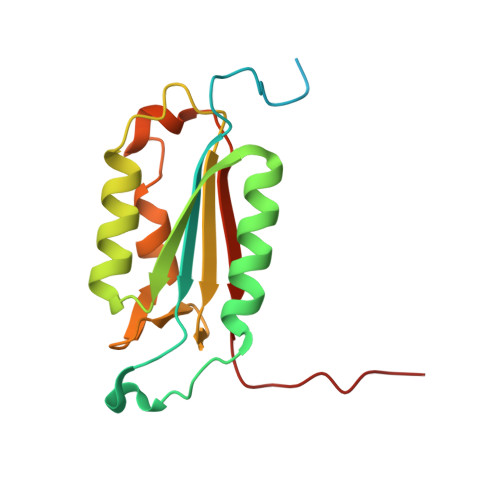
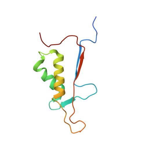
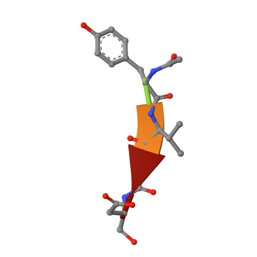
Crystal Structure of Caspase-7 incomplex with Acetyl-YVAD-CHO


Experimental Data Snapshot

  • Method: X-RAY DIFFRACTION
  • Resolution: 2.75 Å
  • R-Value Free: 0.242 
  • R-Value Work: 0.203 
  • R-Value Observed: 0.218 

Starting Model: experimental
View more details

wwPDB Validation   3D Report Full Report


This is version 1.3 of the entry. See complete history


Literature

Conformational similarity in the activation of caspase-3 and -7 revealed by the unliganded and inhibited structures of caspase-7.

Agniswamy, J.Fang, B.Weber, I.T.

(2009) Apoptosis 14: 1135-1144

  • DOI: https://doi.org/10.1007/s10495-009-0388-9
  • Primary Citation of Related Structures:  
    3IBC, 3IBF

  • PubMed Abstract: 

    Caspase-mediated apoptosis has important roles in normal cell differentiation and aging and in many diseases including cancer, neuromuscular disorders and neurodegenerative diseases. Therefore, modulation of caspase activity and conformational states is of therapeutic importance. We report crystal structures of a new unliganded conformation of caspase-7 and the inhibited caspase-7 with the tetrapeptide Ac-YVAD-Cho. Different conformational states and mechanisms for substrate recognition have been proposed based on unliganded structures of the redundant apoptotic executioner caspase-3 and -7. The current study shows that the executioner caspase-3 and -7 have similar conformations for the unliganded active site as well as the inhibitor-bound active site. The new unliganded caspase-7 structure exhibits the tyrosine flipping mechanism in which the Tyr230 has rotated to block entry to the S2 binding site similar to the active site conformation of unliganded caspase-3. The inhibited structure of caspase-7/YVAD shows that the P4 Tyr binds the S4 region specific to polar residues at the expense of a main chain hydrogen bond between the P4 amide and carbonyl oxygen of caspase-7 Gln 276, which is similar to the caspase-3 complex. This new knowledge of the structures and conformational states of unliganded and inhibited caspases will be important for the design of drugs to modulate caspase activity and apoptosis.


  • Organizational Affiliation

    Department of Biology, Georgia State University, Atlanta, GA 30303, USA.


Macromolecules
Find similar proteins by:  (by identity cutoff)  |  3D Structure
Entity ID: 1
MoleculeChains Sequence LengthOrganismDetailsImage
Caspase-7
A, C
173Homo sapiensMutation(s): 0 
Gene Names: CASP7MCH3
EC: 3.4.22.60
UniProt & NIH Common Fund Data Resources
Find proteins for P55210 (Homo sapiens)
Explore P55210 
Go to UniProtKB:  P55210
PHAROS:  P55210
GTEx:  ENSG00000165806 
Entity Groups  
Sequence Clusters30% Identity50% Identity70% Identity90% Identity95% Identity100% Identity
UniProt GroupP55210
Sequence Annotations
Expand
  • Reference Sequence
Find similar proteins by:  (by identity cutoff)  |  3D Structure
Entity ID: 2
MoleculeChains Sequence LengthOrganismDetailsImage
Caspase-7
B, D
97Homo sapiensMutation(s): 0 
Gene Names: CASP7MCH3
EC: 3.4.22.60
UniProt & NIH Common Fund Data Resources
Find proteins for P55210 (Homo sapiens)
Explore P55210 
Go to UniProtKB:  P55210
PHAROS:  P55210
GTEx:  ENSG00000165806 
Entity Groups  
Sequence Clusters30% Identity50% Identity70% Identity90% Identity95% Identity100% Identity
UniProt GroupP55210
Sequence Annotations
Expand
  • Reference Sequence

Find similar proteins by:  Sequence   |   3D Structure  

Entity ID: 3
MoleculeChains Sequence LengthOrganismDetailsImage
Acetyl-YVAD-CHO
E, F
5N/AMutation(s): 0 
Sequence Annotations
Expand
  • Reference Sequence
Experimental Data & Validation

Experimental Data

  • Method: X-RAY DIFFRACTION
  • Resolution: 2.75 Å
  • R-Value Free: 0.242 
  • R-Value Work: 0.203 
  • R-Value Observed: 0.218 
  • Space Group: P 32 2 1
Unit Cell:
Length ( Å )Angle ( ˚ )
a = 88.248α = 90
b = 88.248β = 90
c = 188.15γ = 120
Software Package:
Software NamePurpose
HKL-2000data collection
PHASERphasing
CNSrefinement
HKL-2000data reduction
HKL-2000data scaling

Structure Validation

View Full Validation Report


Entry History 

Deposition Data

Revision History  (Full details and data files)

  • Version 1.0: 2009-09-01
    Type: Initial release
  • Version 1.1: 2011-07-13
    Changes: Version format compliance
  • Version 1.2: 2023-09-06
    Changes: Data collection, Database references, Derived calculations, Refinement description
  • Version 1.3: 2024-11-27
    Changes: Structure summary