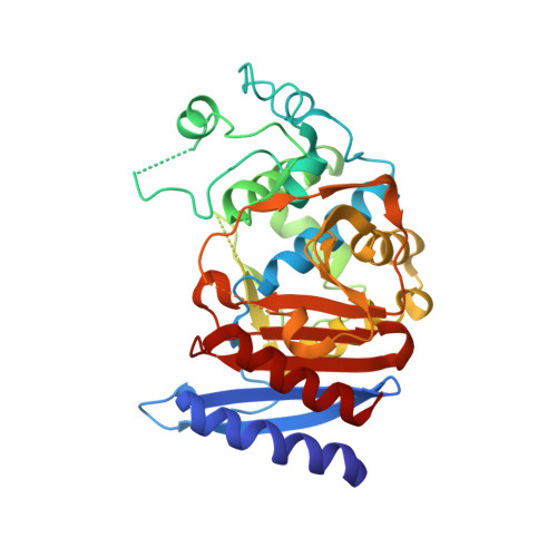
Structural bases for stability-function tradeoffs in antibiotic resistance.
Thomas, V.L., McReynolds, A.C., Shoichet, B.K.(2010) J Mol Biology 396: 47-59
- PubMed: 19913034
- DOI: https://doi.org/10.1016/j.jmb.2009.11.005
- Primary Citation of Related Structures:
3IWI, 3IWO, 3IWQ, 3IXB, 3IXD, 3IXG, 3IXH - PubMed Abstract:
Preorganization of enzyme active sites for substrate recognition typically comes at a cost to the stability of the folded form of the protein; consequently, enzymes can be dramatically stabilized by substitutions that attenuate the size and preorganization "strain" of the active site. How this stability-activity tradeoff constrains enzyme evolution has remained less certain, and it is unclear whether one should expect major stability insults as enzymes mutate towards new activities or how these new activities manifest structurally. These questions are both germane and easy to study in beta-lactamases, which are evolving on the timescale of years to confer resistance to an ever-broader spectrum of beta-lactam antibiotics. To explore whether stability is a substantial constraint on this antibiotic resistance evolution, we investigated extended-spectrum mutants of class C beta-lactamases, which had evolved new activity versus third-generation cephalosporins. Five mutant enzymes had between 100-fold and 200-fold increased activity against the antibiotic cefotaxime in enzyme assays, and the mutant enzymes all lost thermodynamic stability (from 1.7 kcal mol(-)(1) to 4.1 kcal mol(-)(1)), consistent with the stability-function hypothesis. Intriguingly, several of the substitutions were 10-20 A from the catalytic serine; the question of how they conferred extended-spectrum activity arose. Eight structures, including complexes with inhibitors and extended-spectrum antibiotics, were determined by X-ray crystallography. Distinct mechanisms of action, including changes in the flexibility and ground-state structures of the enzyme, are revealed for each mutant. These results explain the structural bases for the antibiotic resistance conferred by these substitutions and their corresponding decrease in protein stability, which will constrain the evolution of new antibiotic resistance.
- Graduate Program in Pharmaceutical Sciences and Pharmacogenomics, University of California San Francisco, San Francisco, CA 94158-2518, USA.
Organizational Affiliation:


