3JT6 | pdb_00003jt6

Structure of neuronal nitric oxide synthase heme domain complexed with N~5~-[4-(methylsulfanyl)butanimidoyl]-L-ornithine


Experimental Data Snapshot

  • Method: X-RAY DIFFRACTION
  • Resolution: 2.20 Å
  • R-Value Free: 
    0.244 (Depositor), 0.236 (DCC) 
  • R-Value Work: 
    0.187 (Depositor) 
  • R-Value Observed: 
    0.190 (Depositor) 

Starting Model: experimental
View more details

wwPDB Validation 3D Report Full Report

Validation slider image for 3JT6

Ligand Structure Quality Assessment 


This is version 1.4 of the entry. See complete history

Literature

Heme-coordinating inhibitors of neuronal nitric oxide synthase. Iron-thioether coordination is stabilized by hydrophobic contacts without increased inhibitor potency.

Martell, J.D.Li, H.Doukov, T.Martasek, P.Roman, L.J.Soltis, M.Poulos, T.L.Silverman, R.B.

(2010) J Am Chem Soc 132: 798-806

  • DOI: https://doi.org/10.1021/ja908544f
  • Primary Citation Related Structures: 
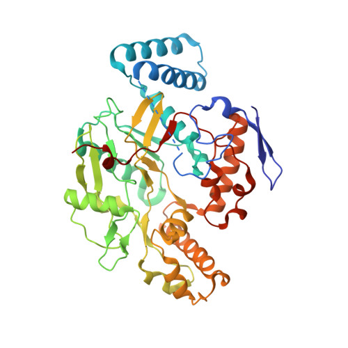
    3JT3, 3JT4, 3JT5, 3JT6, 3JT7, 3JT8, 3JT9, 3JTA

  • PubMed Abstract: 

    The heme-thioether ligand interaction often occurs between heme iron and native methionine ligands, but thioether-based heme-coordinating (type II) inhibitors are uncommon due to the difficulty in stabilizing the Fe-S bond. Here, a thioether-based inhibitor (3) of neuronal nitric oxide synthase (nNOS) was designed, and its binding was characterized by spectrophotometry and crystallography. A crystal structure of inhibitor 3 coordinated to heme iron was obtained, representing, to our knowledge, the first crystal structure of a thioether inhibitor complexed to any heme enzyme. A series of related potential inhibitors (4-8) also were evaluated. Compounds 4-8 were all found to be type I (non-heme-coordinating) inhibitors of ferric nNOS, but 4 and 6-8 were found to switch to type II upon heme reduction to the ferrous state, reflecting the higher affinity of thioethers for ferrous heme than for ferric heme. Contrary to what has been widely thought, thioether-heme ligation was found not to increase inhibitor potency, illustrating the intrinsic weakness of the thioether-ferric heme linkage. Subtle changes in the alkyl groups attached to the thioether sulfur caused drastic changes in the binding conformation, indicating that hydrophobic contacts play a crucial role in stabilizing the thioether-heme coordination.


  • Organizational Affiliation
    • Department of Chemistry, Center for Molecular Innovation and Drug Discovery, and Chemistry of Life Processes Institute, Northwestern University, Evanston, Illinois 60208-3113, USA.

Macromolecule Content 

  • Total Structure Weight: 100.02 kDa 
  • Atom Count: 7,031 
  • Modeled Residue Count: 818 
  • Deposited Residue Count: 844 
  • Unique protein chains: 1

Macromolecules

Find similar proteins by:|  3D Structure
Entity ID: 1
MoleculeChains  Sequence LengthOrganismDetailsImage
Nitric oxide synthase, brain
A, B
422Rattus norvegicusMutation(s): 0 
Gene Names: Nos1Bnos
EC: 1.14.13.39
UniProt
Find proteins for P29476 (Rattus norvegicus)
Explore P29476 
Go to UniProtKB:  P29476
Entity Groups
Sequence Clusters30% Identity50% Identity70% Identity90% Identity95% Identity100% Identity
UniProt GroupP29476
Sequence Annotations
Expand
Reference Sequence

Small Molecules

Ligands 5 Unique
IDChains Name / Formula / InChI Key2D Diagram3D Interactions
HEM

Query on HEM


Download:Ideal Coordinates CCD File
C [auth A],
H [auth B]
PROTOPORPHYRIN IX CONTAINING FE
C34 H32 Fe N4 O4
KABFMIBPWCXCRK-RGGAHWMASA-L
JM6

Query on JM6


Download:Ideal Coordinates CCD File
E [auth A],
J [auth B]
N~5~-[4-(methylsulfanyl)butanimidoyl]-L-ornithine
C10 H21 N3 O2 S
BHXCYHGJTSQWOB-QMMMGPOBSA-N
H4B

Query on H4B


Download:Ideal Coordinates CCD File
D [auth A],
I [auth B]
5,6,7,8-TETRAHYDROBIOPTERIN
C9 H15 N5 O3
FNKQXYHWGSIFBK-RPDRRWSUSA-N
ZN

Query on ZN


Download:Ideal Coordinates CCD File
G [auth A]ZINC ION
Zn
PTFCDOFLOPIGGS-UHFFFAOYSA-N
ACT

Query on ACT


Download:Ideal Coordinates CCD File
F [auth A],
K [auth B]
ACETATE ION
C2 H3 O2
QTBSBXVTEAMEQO-UHFFFAOYSA-M

Experimental Data & Validation

Experimental Data

  • Method: X-RAY DIFFRACTION
  • Resolution: 2.20 Å
  • R-Value Free:  0.244 (Depositor), 0.236 (DCC) 
  • R-Value Work:  0.187 (Depositor) 
  • R-Value Observed: 0.190 (Depositor) 
Space Group: P 21 21 21
Unit Cell:
Length ( Å )Angle ( ˚ )
a = 51.84α = 90
b = 110.77β = 90
c = 164.58γ = 90
Software Package:
Software NamePurpose
REFMACrefinement
CNSrefinement
Blu-Icedata collection
HKL-2000data reduction
SCALEPACKdata scaling
CNSphasing

Structure Validation

View Full Validation Report


Ligand Structure Quality Assessment 


Entry History 

Deposition Data

Revision History  (Full details and data files)

  • Version 1.0: 2010-01-12
    Type: Initial release
  • Version 1.1: 2011-07-13
    Changes: Version format compliance
  • Version 1.2: 2017-11-01
    Changes: Refinement description
  • Version 1.3: 2019-07-24
    Changes: Data collection, Refinement description
  • Version 1.4: 2023-09-06
    Changes: Data collection, Database references, Derived calculations, Refinement description