


Structure of the CBS pair of a putative D-arabinose 5-phosphate isomerase from Klebsiella pneumoniae subsp. pneumoniae.
Cuff, M.E., Volkart, L., Bearden, J., Joachimiak, A.To be published.
Experimental Data Snapshot
Entity ID: 1 | |||||
|---|---|---|---|---|---|
| Molecule | Chains | Sequence Length | Organism | Details | Image |
| putative D-arabinose 5-phosphate isomerase | 149 | Klebsiella pneumoniae subsp. pneumoniae MGH 78578 | Mutation(s): 0 Gene Names: KPN78578_35760, KPN_03607, yrbH EC: 5.3.1.13 | 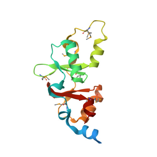 | |
UniProt | |||||
Entity Groups | |||||
| Sequence Clusters | 30% Identity50% Identity70% Identity90% Identity95% Identity100% Identity | ||||
| UniProt Group | A6TEL6 | ||||
Sequence AnnotationsExpand | |||||
Reference Sequence | |||||
| Ligands 3 Unique | |||||
|---|---|---|---|---|---|
| ID | Chains | Name / Formula / InChI Key | 2D Diagram | 3D Interactions | |
| CMK Download:Ideal Coordinates CCD File | C [auth A], G [auth B] | CYTIDINE 5'-MONOPHOSPHATE 3-DEOXY-BETA-D-GULO-OCT-2-ULO-PYRANOSONIC ACID C17 H26 N3 O15 P YWWJKULNWGRYAS-UOVSKDHASA-N |  | ||
| SO4 Download:Ideal Coordinates CCD File | D [auth A] | SULFATE ION O4 S QAOWNCQODCNURD-UHFFFAOYSA-L |  | ||
| GOL Download:Ideal Coordinates CCD File | E [auth A], F [auth A], H [auth B] | GLYCEROL C3 H8 O3 PEDCQBHIVMGVHV-UHFFFAOYSA-N |  | ||
| Modified Residues 1 Unique | |||||
|---|---|---|---|---|---|
| ID | Chains | Type | Formula | 2D Diagram | Parent |
| MSE Query on MSE | A, B | L-PEPTIDE LINKING | C5 H11 N O2 Se |  | MET |
| Length ( Å ) | Angle ( ˚ ) |
|---|---|
| a = 102.151 | α = 90 |
| b = 102.151 | β = 90 |
| c = 214.85 | γ = 120 |
| Software Name | Purpose |
|---|---|
| DENZO | data reduction |
| SCALEPACK | data scaling |
| MLPHARE | phasing |
| DM | phasing |
| REFMAC | refinement |
| PDB_EXTRACT | data extraction |
| SBC-Collect | data collection |
| HKL-3000 | data reduction |
| HKL-3000 | data scaling |
| HKL-3000 | phasing |
| SHELXD | phasing |
| SHELXE | model building |
| SOLVE | phasing |
| RESOLVE | phasing |
| ARP/wARP | model building |
| CCP4 | phasing |
| O | model building |
| Coot | model building |


