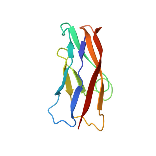
3K6I | pdb_00003k6i

Crystal structure of chicken T-cadherin EC1


Experimental Data Snapshot

  • Method: X-RAY DIFFRACTION
  • Resolution: 1.13 Å
  • R-Value Free: 
    0.158 (Depositor), 0.155 (DCC) 
  • R-Value Work: 
    0.142 (Depositor), 0.151 (DCC) 
  • R-Value Observed: 
    0.143 (Depositor) 

Starting Model: experimental
View more details

wwPDB Validation 3D Report Full Report

Validation slider image for 3K6I

This is version 1.2 of the entry. See complete history. 

Literature

T-cadherin structures reveal a novel adhesive binding mechanism

Ciatto, C., Bahna, F., Zampieri, N., Vansteenhouse, H.C., Katsamba, P.S., Ahlsen, G., Harrison, O.J., Brasch, J., Jin, X., Posy, S., Vendome, J., Ranscht, B., Jessell, T.M., Honig, B., Shapiro, L.

(2010) Nat Struct Mol Biol 17: 339-347

  • DOI: https://doi.org/10.1038/nsmb.1781
  • Primary Citation Related Structures: 
    3K5R, 3K5S, 3K6D, 3K6F, 3K6I

  • PubMed Abstract: 

    Vertebrate genomes encode 19 classical cadherins and about 100 nonclassical cadherins. Adhesion by classical cadherins depends on binding interactions in their N-terminal EC1 domains, which swap N-terminal beta-strands between partner molecules from apposing cells. However, strand-swapping sequence signatures are absent from nonclassical cadherins, raising the question of how these proteins function in adhesion. Here, we show that T-cadherin, a glycosylphosphatidylinositol (GPI)-anchored cadherin, forms dimers through an alternative nonswapped interface near the EC1-EC2 calcium-binding sites. Mutations within this interface ablate the adhesive capacity of T-cadherin. These nonadhesive T-cadherin mutants also lose the ability to regulate neurite outgrowth from T-cadherin-expressing neurons. Our findings reveal the likely molecular architecture of the T-cadherin homophilic interface and its requirement for axon outgrowth regulation. The adhesive binding mode used by T-cadherin may also be used by other nonclassical cadherins.


  • Organizational Affiliation: 
    • Department of Biochemistry and Molecular Biophysics, Columbia University, New York, New York, USA.

Macromolecule Content 

  • Total Structure Weight: 10.93 kDa 
  • Atom Count: 1,117 
  • Modeled Residue Count: 99 
  • Deposited Residue Count: 99 
  • Unique protein chains: 1

Macromolecules

Find similar proteins by:|  3D Structure
Entity ID: 1
MoleculeChains  Sequence LengthOrganismDetailsImage
T-cadherin99Gallus gallusMutation(s): 0 
Gene Names: CDH13
UniProt
Find proteins for P33150 (Gallus gallus)
Explore P33150 
Go to UniProtKB:  P33150
Entity Groups
Sequence Clusters30% Identity50% Identity70% Identity90% Identity95% Identity100% Identity
UniProt GroupP33150
Sequence Annotations
Expand
Reference Sequence

Experimental Data & Validation

Experimental Data

  • Method: X-RAY DIFFRACTION
  • Resolution: 1.13 Å
  • R-Value Free:  0.158 (Depositor), 0.155 (DCC) 
  • R-Value Work:  0.142 (Depositor), 0.151 (DCC) 
  • R-Value Observed: 0.143 (Depositor) 
Space Group: P 21 21 21
Unit Cell:
Length ( Å )Angle ( ˚ )
a = 44.153α = 90
b = 44.93β = 90
c = 56.666γ = 90
Software Package:
Software NamePurpose
REFMACrefinement
PDB_EXTRACTdata extraction
HKL-2000data collection
XDSdata reduction
XSCALEdata scaling
PHASERphasing

Structure Validation

View Full Validation Report


Entry History 

Deposition Data

Revision History  (Full details and data files)

  • Version 1.0: 2010-03-02
    Type: Initial release
  • Version 1.1: 2011-07-13
    Changes: Version format compliance
  • Version 1.2: 2023-09-06
    Changes: Data collection, Database references, Derived calculations, Refinement description