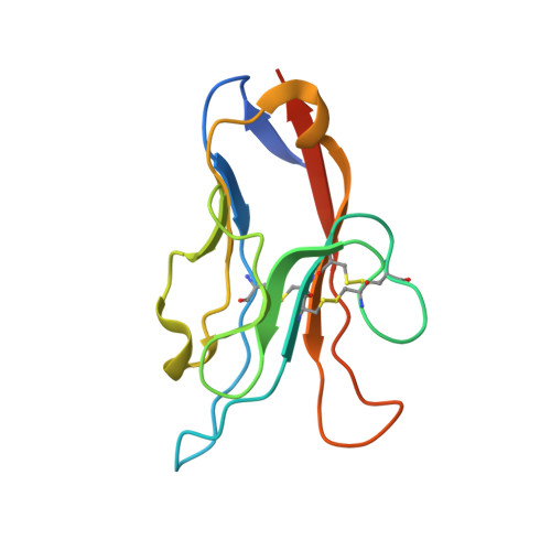
T cell/transmembrane, Ig, and mucin-3 allelic variants differentially recognize phosphatidylserine and mediate phagocytosis of apoptotic cells.
DeKruyff, R.H., Bu, X., Ballesteros, A., Santiago, C., Chim, Y.L., Lee, H.H., Karisola, P., Pichavant, M., Kaplan, G.G., Umetsu, D.T., Freeman, G.J., Casasnovas, J.M.(2010) J Immunol 184: 1918-1930
- PubMed: 20083673
- DOI: https://doi.org/10.4049/jimmunol.0903059
- Primary Citation of Related Structures:
3KAA - PubMed Abstract:
T cell/transmembrane, Ig, and mucin (TIM) proteins, identified using a congenic mouse model of asthma, critically regulate innate and adaptive immunity. TIM-1 and TIM-4 are receptors for phosphatidylserine (PtdSer), exposed on the surfaces of apoptotic cells. Herein, we show with structural and biological studies that TIM-3 is also a receptor for PtdSer that binds in a pocket on the N-terminal IgV domain in coordination with a calcium ion. The TIM-3/PtdSer structure is similar to that of TIM-4/PtdSer, reflecting a conserved PtdSer binding mode by TIM family members. Fibroblastic cells expressing mouse or human TIM-3 bound and phagocytosed apoptotic cells, with the BALB/c allelic variant of mouse TIM-3 showing a higher capacity than the congenic C.D2 Es-Hba-allelic variant. These functional differences were due to structural differences in the BC loop of the IgV domain of the TIM-3 polymorphic variants. In contrast to fibroblastic cells, T or B cells expressing TIM-3 formed conjugates with but failed to engulf apoptotic cells. Together these findings indicate that TIM-3-expressing cells can respond to apoptotic cells, but the consequence of TIM-3 engagement of PtdSer depends on the polymorphic variants of and type of cell expressing TIM-3. These findings establish a new paradigm for TIM proteins as PtdSer receptors and unify the function of the TIM gene family, which has been associated with asthma and autoimmunity and shown to modulate peripheral tolerance.
- Division of Immunology, Children's Hospital Boston, e, Boston, MA, USA.
Organizational Affiliation:


