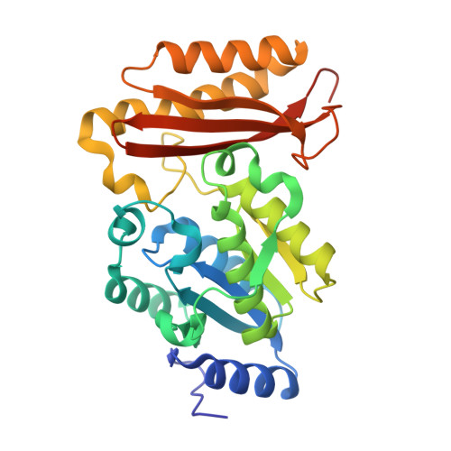
3LE8

Crystal Structure of Mycobacterium Tuberculosis Pantothenate Synthetase at 1.70 Angstrom resolution in complex with 2-(2-((benzofuran-2-carboxamido)methyl)-5-methoxy-1H-indol-1-yl)acetic acid


Experimental Data Snapshot

  • Method: X-RAY DIFFRACTION
  • Resolution: 1.70 Å
  • R-Value Free: 0.223 
  • R-Value Work: 0.172 
  • R-Value Observed: 0.175 

Starting Model: experimental
View more details

wwPDB Validation   3D Report Full Report


Ligand Structure Quality Assessment 


This is version 1.3 of the entry. See complete history


Literature

Optimization of the interligand overhauser effect for fragment linking: application to inhibitor discovery against Mycobacterium tuberculosis pantothenate synthetase.

Sledz, P.Silvestre, H.L.Hung, A.W.Ciulli, A.Blundell, T.L.Abell, C.

(2010) J Am Chem Soc 132: 4544-4545

  • DOI: https://doi.org/10.1021/ja100595u
  • Primary Citation of Related Structures:  
    3LE8

  • PubMed Abstract: 

    Fragment-based methods are a new and emerging approach for the discovery of protein binders that are potential new therapeutic agents. Several ways of utilizing structural information to guide the inhibitor assembly have been explored to date. One of the approaches, application of interligand Overhauser effect (ILOE) observations, is of particular interest, as it does not require the availability of a three-dimensional protein structure and is an NMR-based method that can be applied to targets that cannot be observed directly because of their size. Fragments, as small and often hydrophobic molecules, suffer from problems including compound aggregation in an aqueous environment and nonspecific binding contributions, especially when screened at higher concentrations suitable for ILOE observations. Here we report how this problem can be overcome by applying a step-by-step iterative procedure that includes the application of optimized probe molecules with known binding modes to elucidate the unknown binding modes of fragments. An enzyme substrate with well-characterized binding was used as a starting point, and the relative binding modes of modified fragments derived from ILOE observations were used to guide the fragment linking, leading to a potent inhibitor of our model system, Mycobacterium tuberculosis pantothenate synthetase, a potential drug target. We have supported our NMR data with crystal structures, thus establishing the guidelines for optimizing the ILOE observations. This model study should expand the application of the technique in drug discovery.


  • Organizational Affiliation

    University Chemical Laboratory, University of Cambridge, Lensfield Road, Cambridge CB2 1EW, United Kingdom.


Macromolecules
Find similar proteins by:  (by identity cutoff)  |  3D Structure
Entity ID: 1
MoleculeChains Sequence LengthOrganismDetailsImage
Pantothenate synthetase
A, B
300Mycobacterium tuberculosis H37RvMutation(s): 2 
Gene Names: MT3707MTCY07H7B.20panCRv3602c
EC: 6.3.2.1
UniProt
Find proteins for P9WIL5 (Mycobacterium tuberculosis (strain ATCC 25618 / H37Rv))
Explore P9WIL5 
Go to UniProtKB:  P9WIL5
Entity Groups  
Sequence Clusters30% Identity50% Identity70% Identity90% Identity95% Identity100% Identity
UniProt GroupP9WIL5
Sequence Annotations
Expand
  • Reference Sequence
Small Molecules
Ligands 4 Unique
IDChains Name / Formula / InChI Key2D Diagram3D Interactions
2B5
Query on 2B5

Download Ideal Coordinates CCD File 
C [auth A],
K [auth B]
2-(2-((benzofuran-2-carboxamido)methyl)-5-methoxy-1H-indol-1-yl)acetic acid
C21 H18 N2 O5
CYMXZVQAGCGDHU-UHFFFAOYSA-N
GOL
Query on GOL

Download Ideal Coordinates CCD File 
F [auth A],
L [auth B],
M [auth B],
N [auth B]
GLYCEROL
C3 H8 O3
PEDCQBHIVMGVHV-UHFFFAOYSA-N
EDO
Query on EDO

Download Ideal Coordinates CCD File 
D [auth A],
E [auth A],
G [auth A],
H [auth A],
J [auth A]
1,2-ETHANEDIOL
C2 H6 O2
LYCAIKOWRPUZTN-UHFFFAOYSA-N
EOH
Query on EOH

Download Ideal Coordinates CCD File 
I [auth A]ETHANOL
C2 H6 O
LFQSCWFLJHTTHZ-UHFFFAOYSA-N
Binding Affinity Annotations 
IDSourceBinding Affinity
2B5 BindingDB:  3LE8 Kd: 860 (nM) from 1 assay(s)
PDBBind:  3LE8 Kd: 860 (nM) from 1 assay(s)
Experimental Data & Validation

Experimental Data

  • Method: X-RAY DIFFRACTION
  • Resolution: 1.70 Å
  • R-Value Free: 0.223 
  • R-Value Work: 0.172 
  • R-Value Observed: 0.175 
  • Space Group: P 1 21 1
Unit Cell:
Length ( Å )Angle ( ˚ )
a = 48.58α = 90
b = 71.14β = 99.04
c = 81.49γ = 90
Software Package:
Software NamePurpose
AMoREphasing
REFMACrefinement
MOSFLMdata reduction
SCALAdata scaling

Structure Validation

View Full Validation Report


Ligand Structure Quality Assessment 


Entry History 

Deposition Data

Revision History  (Full details and data files)

  • Version 1.0: 2010-04-14
    Type: Initial release
  • Version 1.1: 2011-07-13
    Changes: Version format compliance
  • Version 1.2: 2021-10-13
    Changes: Database references, Derived calculations
  • Version 1.3: 2023-09-06
    Changes: Data collection, Refinement description