3LXE | pdb_00003lxe

Human Carbonic Anhydrase I in complex with topiramate


Experimental Data Snapshot

  • Method: X-RAY DIFFRACTION
  • Resolution: 1.90 Å
  • R-Value Free: 
    0.228 (Depositor), 0.228 (DCC) 
  • R-Value Work: 
    0.199 (Depositor), 0.199 (DCC) 

Starting Model: experimental
View more details

wwPDB Validation 3D Report Full Report

Validation slider image for 3LXE

Ligand Structure Quality Assessment 


This is version 1.2 of the entry. See complete history

Literature

The first example of a significant active site conformational rearrangement in a carbonic anhydrase-inhibitor adduct: the carbonic anhydrase I-topiramate complex.

Alterio, V.Monti, S.M.Truppo, E.Pedone, C.Supuran, C.T.De Simone, G.

(2010) Org Biomol Chem 

  • DOI: https://doi.org/10.1039/b926832d
  • Primary Citation Related Structures: 
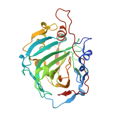
    3LXE

  • PubMed Abstract: 

    Topiramate is a widely used antiepileptic drug, which has been demonstrated to act as an efficient weight loss agent. Since several studies have pointed out that is a potent in vitro inhibitor of several Carbonic anhydrase (CA) isozymes, it has been hypothesized that its anti-obesity properties could be ascribed to the inhibition of the CAs involved in de novo lipogenesis. Consequently, the study of the interactions of with all human CA isoforms represents an important step for the rational drug design of selective CA inhibitors to be used as anti-obesity drugs. In this paper we report the crystallographic structure of the adduct that forms with hCA I, showing for the first time a profound reorganization of the CA active site upon binding of the inhibitor. Moreover, a structural comparison with hCA II- and hCA VA- adducts, previously investigated, has been performed showing that a different H-bond network together with the movement of some amino acid residues in the active site may account for the different inhibition constants of toward these three CA isozymes.


  • Organizational Affiliation
    • Istituto di Biostrutture e Bioimmagini-CNR, via Mezzocannone 16, 80134 Naples, Italy.

Macromolecule Content 

  • Total Structure Weight: 58.36 kDa 
  • Atom Count: 4,532 
  • Modeled Residue Count: 512 
  • Deposited Residue Count: 520 
  • Unique protein chains: 1

Macromolecules

Find similar proteins by:|  3D Structure
Entity ID: 1
MoleculeChains  Sequence LengthOrganismDetailsImage
Carbonic anhydrase 1
A, B
260Homo sapiensMutation(s): 0 
EC: 4.2.1.1 (PDB Primary Data), 4.2.1.69 (UniProt)
UniProt & NIH Common Fund Data Resources
Find proteins for P00915 (Homo sapiens)
Explore P00915 
Go to UniProtKB:  P00915
PHAROS:  P00915
GTEx:  ENSG00000133742 
Entity Groups
Sequence Clusters30% Identity50% Identity70% Identity90% Identity95% Identity100% Identity
UniProt GroupP00915
Sequence Annotations
Expand
Reference Sequence

Experimental Data & Validation

Experimental Data

  • Method: X-RAY DIFFRACTION
  • Resolution: 1.90 Å
  • R-Value Free:  0.228 (Depositor), 0.228 (DCC) 
  • R-Value Work:  0.199 (Depositor), 0.199 (DCC) 
Space Group: P 21 21 21
Unit Cell:
Length ( Å )Angle ( ˚ )
a = 63.32α = 90
b = 71.07β = 90
c = 120.66γ = 90
Software Package:
Software NamePurpose
HKL-2000data collection
AMoREphasing
CNSrefinement
HKL-2000data reduction
HKL-2000data scaling

Structure Validation

View Full Validation Report


Ligand Structure Quality Assessment 


Entry History 

Deposition Data

Revision History  (Full details and data files)

  • Version 1.0: 2010-07-21
    Type: Initial release
  • Version 1.1: 2011-07-13
    Changes: Version format compliance
  • Version 1.2: 2023-11-01
    Changes: Data collection, Database references, Derived calculations, Refinement description