3MAW

Structure of the Newcastle disease virus F protein in the post-fusion conformation


Experimental Data Snapshot

  • Method: X-RAY DIFFRACTION
  • Resolution: 3.50 Å
  • R-Value Free: 
    0.294 (Depositor), 0.284 (DCC) 
  • R-Value Work: 
    0.261 (Depositor), 0.251 (DCC) 
  • R-Value Observed: 
    0.263 (Depositor) 

wwPDB Validation   3D Report Full Report


Ligand Structure Quality Assessment 

Created with Raphaël 2.3.0Worse 01 BetterLigand structure goodness of fit to experimental dataBest fitted NDGClick on this verticalbar to view details

This is version 2.1 of the entry. See complete history


Literature

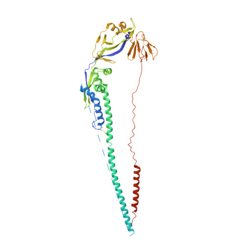
Structure of the Newcastle disease virus F protein in the post-fusion conformation.

Swanson, K.Wen, X.Leser, G.P.Paterson, R.G.Lamb, R.A.Jardetzky, T.S.

(2010) Virology 402: 372-379

  • DOI: https://doi.org/10.1016/j.virol.2010.03.050
  • Primary Citation of Related Structures:  
    3MAW

  • PubMed Abstract: 

    The paramyxovirus F protein is a class I viral membrane fusion protein which undergoes a significant refolding transition during virus entry. Previous studies of the Newcastle disease virus, human parainfluenza virus 3 and parainfluenza virus 5 F proteins revealed differences in the pre- and post-fusion structures. The NDV Queensland (Q) F structure lacked structural elements observed in the other two structures, which are key to the refolding and fusogenic activity of F. Here we present the NDV Australia-Victoria (AV) F protein post-fusion structure and provide EM evidence for its folding to a pre-fusion form. The NDV AV F structure contains heptad repeat elements missing in the previous NDV Q F structure, forming a post-fusion six-helix bundle (6HB) similar to the post-fusion hPIV3 F structure. Electrostatic and temperature factor analysis of the F structures points to regions of these proteins that may be functionally important in their membrane fusion activity.


  • Organizational Affiliation

    Howard Hughes Medical Institute, USA.


Macromolecules
Find similar proteins by:  (by identity cutoff)  |  3D Structure
Entity ID: 1
MoleculeChains Sequence LengthOrganismDetailsImage
Fusion glycoprotein F0
A, B
483Newcastle disease virus (STRAIN AUSTRALIA-VICTORIA/32)Mutation(s): 0 
Gene Names: F
UniProt
Find proteins for P12572 (Newcastle disease virus (strain Chicken/Australia-Victoria/32))
Explore P12572 
Go to UniProtKB:  P12572
Entity Groups  
Sequence Clusters30% Identity50% Identity70% Identity90% Identity95% Identity100% Identity
UniProt GroupP12572
Glycosylation
Glycosylation Sites: 2
Sequence Annotations
Expand
  • Reference Sequence
Oligosaccharides

Help

Entity ID: 2
MoleculeChains Length2D Diagram Glycosylation3D Interactions
2-acetamido-2-deoxy-beta-D-glucopyranose-(1-4)-2-acetamido-2-deoxy-beta-D-glucopyranose
C, D, E, F
2N-Glycosylation
Glycosylation Resources
GlyTouCan:  G42666HT
GlyCosmos:  G42666HT
GlyGen:  G42666HT
Experimental Data & Validation

Experimental Data

  • Method: X-RAY DIFFRACTION
  • Resolution: 3.50 Å
  • R-Value Free:  0.294 (Depositor), 0.284 (DCC) 
  • R-Value Work:  0.261 (Depositor), 0.251 (DCC) 
  • R-Value Observed: 0.263 (Depositor) 
Space Group: H 3
Unit Cell:
Length ( Å )Angle ( ˚ )
a = 83.253α = 90
b = 83.253β = 90
c = 461.406γ = 120
Software Package:
Software NamePurpose
MAR345dtbdata collection
PHASERphasing
PHENIXrefinement
HKL-2000data reduction
SCALEPACKdata scaling

Structure Validation

View Full Validation Report


Ligand Structure Quality Assessment 

Created with Raphaël 2.3.0Worse 01 BetterLigand structure goodness of fit to experimental dataBest fitted NDGClick on this verticalbar to view details

Entry History 

Deposition Data

Revision History  (Full details and data files)

  • Version 1.0: 2010-05-26
    Type: Initial release
  • Version 1.1: 2011-07-13
    Changes: Version format compliance
  • Version 2.0: 2020-07-29
    Type: Remediation
    Reason: Carbohydrate remediation
    Changes: Advisory, Atomic model, Data collection, Database references, Derived calculations, Structure summary
  • Version 2.1: 2024-11-06
    Changes: Data collection, Database references, Structure summary