3MJX

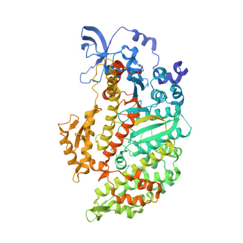
Crystal structure of myosin-2 motor domain in complex with ADP-Metavanadate and blebbistatin


Experimental Data Snapshot

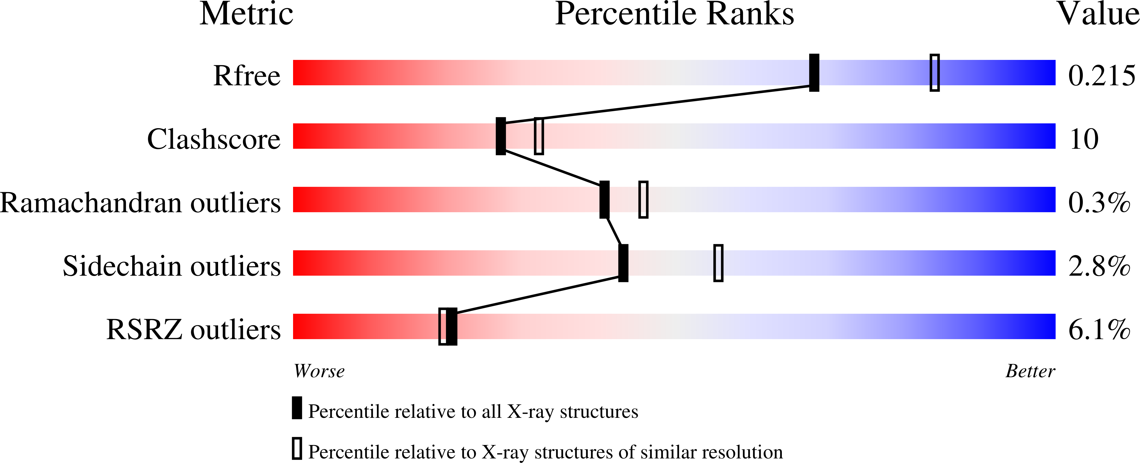
  • Method: X-RAY DIFFRACTION
  • Resolution: 2.20 Å
  • R-Value Free: 0.226 
  • R-Value Work: 0.204 
  • R-Value Observed: 0.205 

Starting Model: experimental
View more details

wwPDB Validation   3D Report Full Report


Ligand Structure Quality Assessment 


This is version 1.3 of the entry. See complete history


Literature

The mechanism of pentabromopseudilin inhibition of myosin motor activity.

Fedorov, R.Bohl, M.Tsiavaliaris, G.Hartmann, F.K.Taft, M.H.Baruch, P.Brenner, B.Martin, R.Knolker, H.J.Gutzeit, H.O.Manstein, D.J.

(2009) Nat Struct Mol Biol 16: 80-88

  • DOI: https://doi.org/10.1038/nsmb.1542
  • Primary Citation of Related Structures:  
    2JHR, 2JJ9, 3MJX

  • PubMed Abstract: 

    We have identified pentabromopseudilin (PBP) as a potent inhibitor of myosin-dependent processes such as isometric tension development and unloaded shortening velocity. PBP-induced reductions in the rate constants for ATP binding, ATP hydrolysis and ADP dissociation extend the time required per myosin ATPase cycle in the absence and presence of actin. Additionally, coupling between the actin and nucleotide binding sites is reduced in the presence of the inhibitor. The selectivity of PBP differs from that observed with other myosin inhibitors. To elucidate the binding mode of PBP, we crystallized the Dictyostelium myosin-2 motor domain in the presence of Mg(2+)-ADP-meta-vanadate and PBP. The electron density for PBP is unambiguous and shows PBP to bind at a previously unknown allosteric site near the tip of the 50-kDa domain, at a distance of 16 A from the nucleotide binding site and 7.5 A away from the blebbistatin binding pocket.


  • Organizational Affiliation

    Research Centre for Structure Analysis, OE8830, Hannover Medical School, 30623 Hannover, Germany.


Macromolecules
Find similar proteins by:  (by identity cutoff)  |  3D Structure
Entity ID: 1
MoleculeChains Sequence LengthOrganismDetailsImage
Myosin-2 heavy chain788Dictyostelium discoideumMutation(s): 0 
UniProt
Find proteins for P08799 (Dictyostelium discoideum)
Explore P08799 
Go to UniProtKB:  P08799
Entity Groups  
Sequence Clusters30% Identity50% Identity70% Identity90% Identity95% Identity100% Identity
UniProt GroupP08799
Sequence Annotations
Expand
  • Reference Sequence
Experimental Data & Validation

Experimental Data

  • Method: X-RAY DIFFRACTION
  • Resolution: 2.20 Å
  • R-Value Free: 0.226 
  • R-Value Work: 0.204 
  • R-Value Observed: 0.205 
  • Space Group: C 2 2 21
Unit Cell:
Length ( Å )Angle ( ˚ )
a = 89.308α = 90
b = 147.026β = 90
c = 154.594γ = 90
Software Package:
Software NamePurpose
MxCuBEdata collection
AMoREphasing
CNSrefinement
XDSdata reduction
XSCALEdata scaling

Structure Validation

View Full Validation Report


Ligand Structure Quality Assessment 


Entry History 

Deposition Data

Revision History  (Full details and data files)

  • Version 1.0: 2011-04-27
    Type: Initial release
  • Version 1.1: 2011-07-13
    Changes: Version format compliance
  • Version 1.2: 2023-09-06
    Changes: Data collection, Database references, Derived calculations, Refinement description, Structure summary
  • Version 1.3: 2024-03-13
    Changes: Source and taxonomy