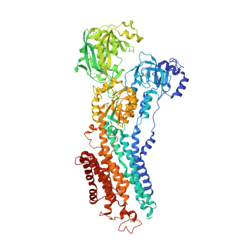
3N5K

Structure Of The (Sr)Ca2+-ATPase E2-AlF4- Form


Experimental Data Snapshot

  • Method: X-RAY DIFFRACTION
  • Resolution: 2.20 Å
  • R-Value Free: 0.216 
  • R-Value Work: 0.184 
  • R-Value Observed: 0.186 

Starting Model: experimental
View more details

wwPDB Validation   3D Report Full Report


Ligand Structure Quality Assessment 


This is version 1.5 of the entry. See complete history


Literature

Ion pathways in the sarcoplasmic reticulum Ca2+-ATPase.

Bublitz, M.Musgaard, M.Poulsen, H.Thogersen, L.Olesen, C.Schiott, B.Morth, J.P.Moller, J.V.Nissen, P.

(2013) J Biol Chem 288: 10759-10765

  • DOI: https://doi.org/10.1074/jbc.R112.436550
  • Primary Citation of Related Structures:  
    3N5K, 3N8G

  • PubMed Abstract: 

    The sarco/endoplasmic reticulum Ca(2+)-ATPase (SERCA) is a transmembrane ion transporter belonging to the P(II)-type ATPase family. It performs the vital task of re-sequestering cytoplasmic Ca(2+) to the sarco/endoplasmic reticulum store, thereby also terminating Ca(2+)-induced signaling such as in muscle contraction. This minireview focuses on the transport pathways of Ca(2+) and H(+) ions across the lipid bilayer through SERCA. The ion-binding sites of SERCA are accessible from either the cytoplasm or the sarco/endoplasmic reticulum lumen, and the Ca(2+) entry and exit channels are both formed mainly by rearrangements of four N-terminal transmembrane α-helices. Recent improvements in the resolution of the crystal structures of rabbit SERCA1a have revealed a hydrated pathway in the C-terminal transmembrane region leading from the ion-binding sites to the cytosol. A comparison of different SERCA conformations reveals that this C-terminal pathway is exclusive to Ca(2+)-free E2 states, suggesting that it may play a functional role in proton release from the ion-binding sites. This is in agreement with molecular dynamics simulations and mutational studies and is in striking analogy to a similar pathway recently described for the related sodium pump. We therefore suggest a model for the ion exchange mechanism in P(II)-ATPases including not one, but two cytoplasmic pathways working in concert.


  • Organizational Affiliation

    Centre for Membrane Pumps in Cells and Disease (PUMPkin), Aarhus University, DK-8000 Aarhus C, Denmark.


Macromolecules
Find similar proteins by:  (by identity cutoff)  |  3D Structure
Entity ID: 1
MoleculeChains Sequence LengthOrganismDetailsImage
Sarcoplasmic/endoplasmic reticulum calcium ATPase 1
A, B
994Oryctolagus cuniculusMutation(s): 0 
EC: 3.6.3.8 (PDB Primary Data), 7.2.2.10 (UniProt)
Membrane Entity: Yes 
UniProt
Find proteins for P04191 (Oryctolagus cuniculus)
Explore P04191 
Go to UniProtKB:  P04191
Entity Groups  
Sequence Clusters30% Identity50% Identity70% Identity90% Identity95% Identity100% Identity
UniProt GroupP04191
Sequence Annotations
Expand
  • Reference Sequence
Small Molecules
Ligands 5 Unique
IDChains Name / Formula / InChI Key2D Diagram3D Interactions
TG1
Query on TG1

Download Ideal Coordinates CCD File 
C [auth A],
I [auth B]
OCTANOIC ACID [3S-[3ALPHA, 3ABETA, 4ALPHA, 6BETA, 6ABETA, 7BETA, 8ALPHA(Z), 9BALPHA]]-6-(ACETYLOXY)-2,3,-3A,4,5,6,6A,7,8,9B-DECAHYDRO-3,3A-DIHYDROXY-3,6,9-TRIMETHYL-8-[(2-METHYL-1-OXO-2-BUTENYL)OX Y]-2-OXO-4-(1-OXOBUTOXY)-AZULENO[4,5-B]FURAN-7-YL ESTER
C34 H50 O12
IXFPJGBNCFXKPI-FSIHEZPISA-N
ALF
Query on ALF

Download Ideal Coordinates CCD File 
E [auth A],
K [auth B]
TETRAFLUOROALUMINATE ION
Al F4
UYOMQIYKOOHAMK-UHFFFAOYSA-J
ACT
Query on ACT

Download Ideal Coordinates CCD File 
G [auth A],
H [auth A],
M [auth B],
N [auth B]
ACETATE ION
C2 H3 O2
QTBSBXVTEAMEQO-UHFFFAOYSA-M
K
Query on K

Download Ideal Coordinates CCD File 
F [auth A],
L [auth B]
POTASSIUM ION
K
NPYPAHLBTDXSSS-UHFFFAOYSA-N
MG
Query on MG

Download Ideal Coordinates CCD File 
D [auth A],
J [auth B]
MAGNESIUM ION
Mg
JLVVSXFLKOJNIY-UHFFFAOYSA-N
Binding Affinity Annotations 
IDSourceBinding Affinity
TG1 PDBBind:  3N5K IC50: 0.2 (nM) from 1 assay(s)
Experimental Data & Validation

Experimental Data

  • Method: X-RAY DIFFRACTION
  • Resolution: 2.20 Å
  • R-Value Free: 0.216 
  • R-Value Work: 0.184 
  • R-Value Observed: 0.186 
  • Space Group: P 21 21 21
Unit Cell:
Length ( Å )Angle ( ˚ )
a = 101.93α = 90
b = 109.42β = 90
c = 276.092γ = 90
Software Package:
Software NamePurpose
remdaq.pilatusdata collection
PHASERphasing
PHENIXrefinement
MOSFLMdata reduction
SCALAdata scaling

Structure Validation

View Full Validation Report


Ligand Structure Quality Assessment 


Entry History 

Deposition Data

Revision History  (Full details and data files)

  • Version 1.0: 2011-06-08
    Type: Initial release
  • Version 1.1: 2011-07-13
    Changes: Version format compliance
  • Version 1.2: 2013-02-27
    Changes: Database references
  • Version 1.3: 2014-03-19
    Changes: Refinement description
  • Version 1.4: 2023-09-06
    Changes: Data collection, Database references, Derived calculations, Refinement description, Structure summary
  • Version 1.5: 2024-11-06
    Changes: Structure summary